3449
Quantitative Magnetic Resonance Imaging Assessment of Cervical Spinal Cord Injury in Rats1Biophysics, Medical College of Wisconsin, Milwaukee, WI, United States, 2Neurosurgery, Medical College of Wisconsin, Milwaukee, WI, United States
Synopsis
This work aims to investigate the relationship neurological outcomes from a cervical contusion spinal cord injury model in rats with early MRI markers including edema, hemorrhage, and diffusion weighted imaging. A rat-specific MRI template was constructed from a histological atlas to permit inter-subject registration. Compared to diffusion tensor imaging (DTI) metrics, filtered-DWI diminished the confounding effects of vasogenic edema and improved depiction of the acute damage. Collectively, the registration pipeline facilitated analysis, and the results support the role of MRI in preclinical assessment of cervical SCI.
Introduction
Magnetic resonance imaging (MRI) has shown promise in predicting long-term functional outcome of spinal cord injury (SCI). While current quantitative assessment involves measuring the volume of edema and hemorrhage, these measurements lack specific biological information. Meanwhile, diffusion weighted magnetic resonance imaging (DWI) captures microstructural changes, and filtered-DWI (fDWI) tailored for the spinal cord enhances detection of axonal damage1. Since cervical SCI is the most common injury in humans2, this work sought to investigate quantitative MRI parameters in a unilateral and bilateral injury model. Quantitative measures of edema (length), hemorrhage (length), and axonal injury (AD, fADC||) were evaluated on T2-weighted, T2*-weighted, DTI and fDWI, respectively. A custom template was created from a rat histologic reference atlas to facilitate inter-subject/modality registration of MRI data and analysis using spinal cord toolbox(SCT)3.Methods
Animals: Forty-three Sprague-Dawley rats received unilateral or bilateral cervical (C5) contusion injury. Using an NYU-MASCIC impactor, a 10-gram weight with a 1.5 diameter tip was dropped from a height of 25 mm at C5. The rats underwent MRI scan at 1 post injury (dpi).Functional Assessment: Motor function was evaluated at 2 dpi and weekly thereafter up to 28 dpi using forelimb locomotor assessment scale (FLAS)4.
Magnetic Resonance Imaging Acquisition: MRI was performed on a 9.4T with a volume coil. Sagittal T2w images were acquired with TR=2000ms, TE=19, 57, & 95ms, NEX=2, RARE factor=4, plane resolution=0.117mm, thickness=0.5mm. Axial T2w images were acquired with TR=2000ms, TE=20, 59 & 99ms, NEX=2, RARE factor=4, in-plane resolution=0.129mm, thickness=1mm. T2*w images were obtained using a 3D isotropic multi-gradient echo with TR/TE=65/2.5, 6 echoes, FA=14, resolution=0.224 mm3. Axial DTI was acquired using 4-shot EPI and TR/TE=1800/28ms, NEX=4, in-plane resolution=0.129mm, slice thickness=1mm, b=800s/mm2, 15 directions. fDWI scheme had identical imaging parameters but differed in its b-values and directions: bfilter(Left-Right)= 2000s/mm2, bprobe(Rostral-Caudal)=0-1000s/mm2 in 10 steps.
Custom Template Generation: The Watson, Paxinos and Kayalioglu spinal cord atlas5 was digitized and manually segmented to white and gray matter (WM, GM). Symmetry was enforced by mutual registration of mirror images of the same section followed by averaging, and the centerline was enforced along the central canal for all sections. Images were stacked and resampled to contain the entire spinal cord in physically realistic dimensions. Images with pseudo T2-weighted contrast were used for MRI registration.
Data Analysis: Axial and sagittal T2w images with highest WM/GM contrast were registered using SCT. Cord levels were manually defined from C2-C7 to achieve z-level alignment, straightening, and in-plane (x-y) image registration in a single step. The registered T2w images were further iteratively registered to the group mean T2w through 5 iterations. Diffusion images were registered to the T2w images in native space to first minimize distortion and were subsequently registered to template space. Edema and hemorrhage length were manually measured at the center slice of sagittal T2w and T2*w images. AD and fADC|| were calculated using registered diffusion images and a C5 whole cord mask. Pearson’s correlation coefficient was calculated between each parameter at 1 dpi and FLAS at 28 dpi.
Results
T2w, T2*w and diffusion images showed the extent of cord damage in both models (Fig. 1). Mean diffusivity (MD) showed clear evidence vasogenic edema (blue triangle; increased MD) and cytotoxic edema (red triangle; decreased MD) at the epicenter. Axonal damage/cytotoxic edema was more clearly visualized in fADC|| (white triangle; decreased fADC||), but vasogenic edema was not evident in the same region as it appeared in MD. Compared to edema, axonal damage was localized to the WM.T2w images were straightened and successfully registered to the digitized atlas of spinal cord (Fig. 2). The average image of all-subjects registration clearly delineates WM and GM, allowing registration across imaging modalities.
Cervical contusion injury at C5 resulted in a mildly impaired forelimb function, especially in the forepaw. The motor function was the lowest at 2 dpi and rapidly recovered within 2 weeks of injury (Fig. 3). The motor function was worse in the bilateral injury than hemi-contusion injury. Edema length was negatively correlated with chronic FLAS score, whereas hemorrhage, AD and fADC|| had no correlation.
Discussion
Qualitatively, the fDWI (fADC||) achieves its intended goal of reducing the confounding effects of vasogenic (extracellular) edema that complicates interpretation of DTI measures, and the region of damage is readily evident on the fADC|| maps. Although DTI maps generally have similar information, it was challenging to appreciate the discrete region of damage.Compared to the brain, spatial registration of the spinal cord has been challenged, and SCT enhanced the accessibility of specialized methods to overcome these limitations for the human spinal cord. Similar methods have not been widely applied to the rodent spinal given its unique challenges. Notably, the rodent cord contains much less CSF which complicates segmentation. The availability of a template will facilitate future efforts for voxel-wise mapping and statistics of rodent spinal cord MRI studies.
The primary limitation of the current study is the low degree of variance in neurological outcomes due to the single severity of injury, which potentially limits the correlation analysis. Sham animals were not included since this can drive artifactual correlations given the ceiling effects and non-linearity of the of behavioral tests.
Acknowledgements
This work was supported by funding from the National Institutes of Neurological Disorders and Stroke (R01NS109090). The authors thank Natasha Wilkins, Matthew Runquist and Qian (Kathleen) Yin for experimental assistance.References
1. Skinner, N. P. et al. Filter-Probe Diffusion Imaging Improves Spinal Cord Injury Outcome Prediction. Ann. Neurol. (2018) doi:10.1002/ana.25260.
2. Cotton, B. A. et al. Respiratory complications and mortality risk associated with thoracic spine injury. J. Trauma 59, 1400–1407; discussion 1407-1409 (2005).
3. De Leener, B. et al. SCT: Spinal Cord Toolbox, an open-source software for processing spinal cord MRI data. NeuroImage 145, 24–43 (2017).
4. Anderson, K. D. et al. Forelimb locomotor assessment scale (FLAS): novel assessment of forelimb dysfunction after cervical spinal cord injury. Exp. Neurol. 220, 23–33 (2009).
5. Watson, C., Paxinos, G., Kayalioglu, G. & Heise, C. Chapter 15 - Atlas of the Rat Spinal Cord. in The Spinal Cord (eds. Watson, C., Paxinos, G. & Kayalioglu, G.) 238–306 (Academic Press, 2009). doi:10.1016/B978-0-12-374247-6.50019-5.
Figures
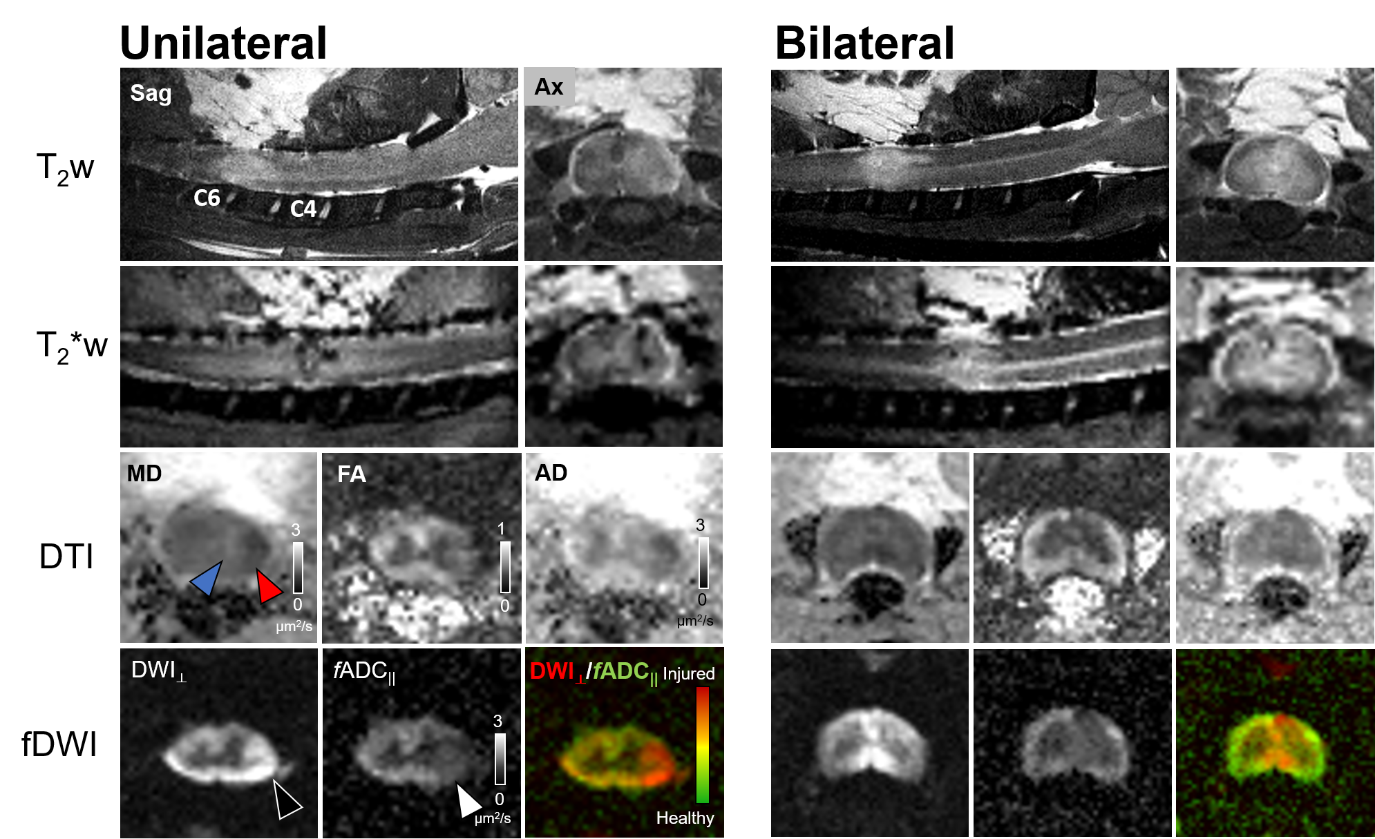
Figure 1 Multi-contrast MRI in acute cervical cord contusion
The extent of the cord damage is captured in different modalities in both unilateral- and bilateral contusion injury. Edema (hyperintensity, T2w), hemorrhage (hypointensity, T2*w), vasogenic edema (blue, black triangle), and cytotoxic edema (red, white triangle) are evident in their respective contrasts. fDWI images clearly delineates focal axonal damage compared to T2w, T2*w and DTI.
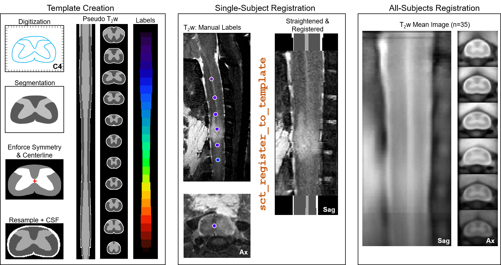
Figure 2 Inter-subject registration pipeline
A histologic spinal cord atlas was digitized across all spinal cord levels to generate a template with pseudo T2-weighted contrast used for MRI registration. Manually marked spinal cord level aided initial z-level alignment and straightening followed by x-y plane registration to the template. Registered single subject images were averaged and registered iteratively across all subjects with the group mean image showing clear spinal cord anatomy.
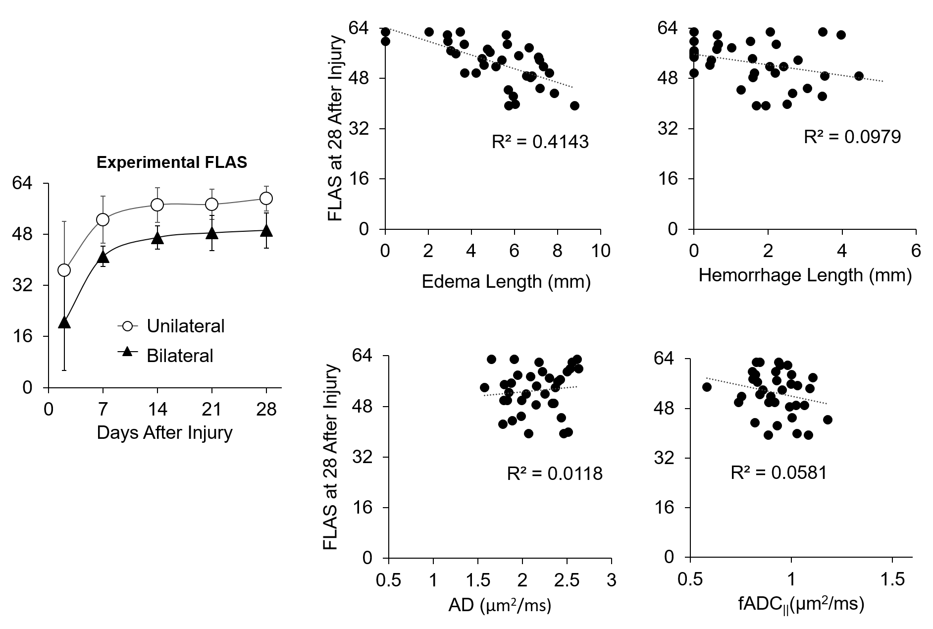
Figure 3 Correlation between acute MRI metrics and chronic motor function (FLAS).
Both injury models showed poor motor function acutely with rapid recovery within 14 days. Quantitative MRI metrics were linearly correlated with long-term functional outcome. Edema length was negatively correlated, while other metrics showed no strong correlations.