3444
Interpreting a machine learning model: radiomics in cervical spondylotic myelopathy postoperative recovery prediction1Peking University Third Hospital, Beijing, China
Synopsis
Previous studies have confirmed that conventional MRI parameters lack stability, and the evaluation of the prognosis of CSM sometimes is controversial. In our study, we first introduced radiomics, a quantitative analysis of image features, into the study of CSM and obtained a reliable and stable model. By analysis features' importance and unboxing the extremely randomized trees model, we came up with assumptions of the relationship between specific features and post-surgical recovery prediction.
Object
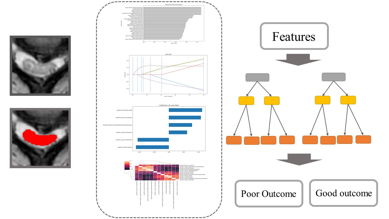
Radiomics, the computerized high-throughput extraction of image features or textures from non-invasive medical imaging, makes it possible for physicians to describe lesions objectively and quantitatively. We developed and validated a radiomic featured base extremely randomized trees model for the prediction of postoperative recovery in cervical spondylotic myelopathy (CSM). The extremely randomized trees model is an ensemble method, which combines several independent estimators to reduce variance and improve robustness.Methods
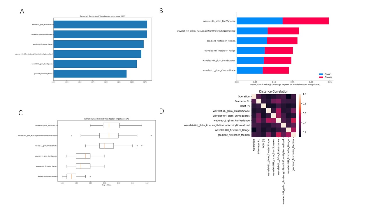
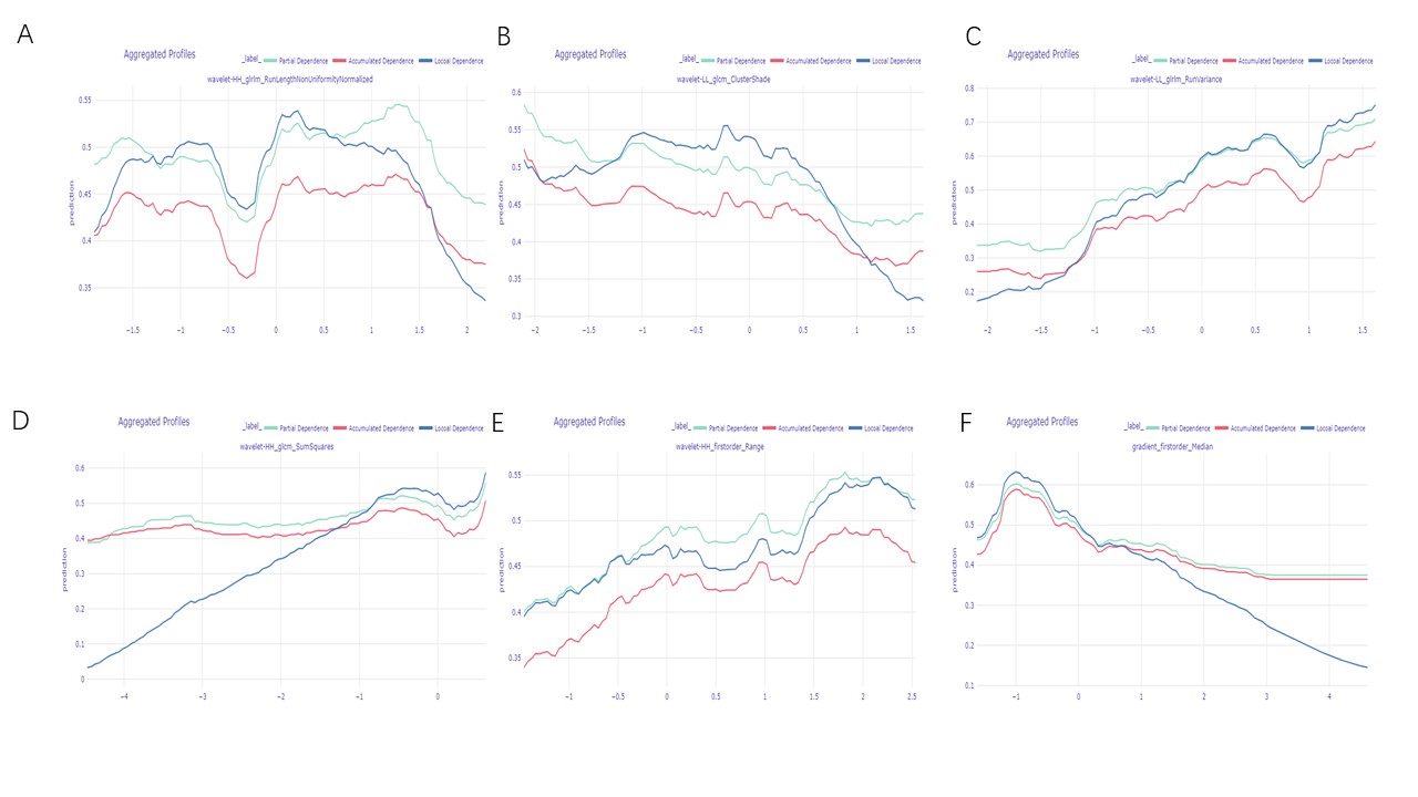
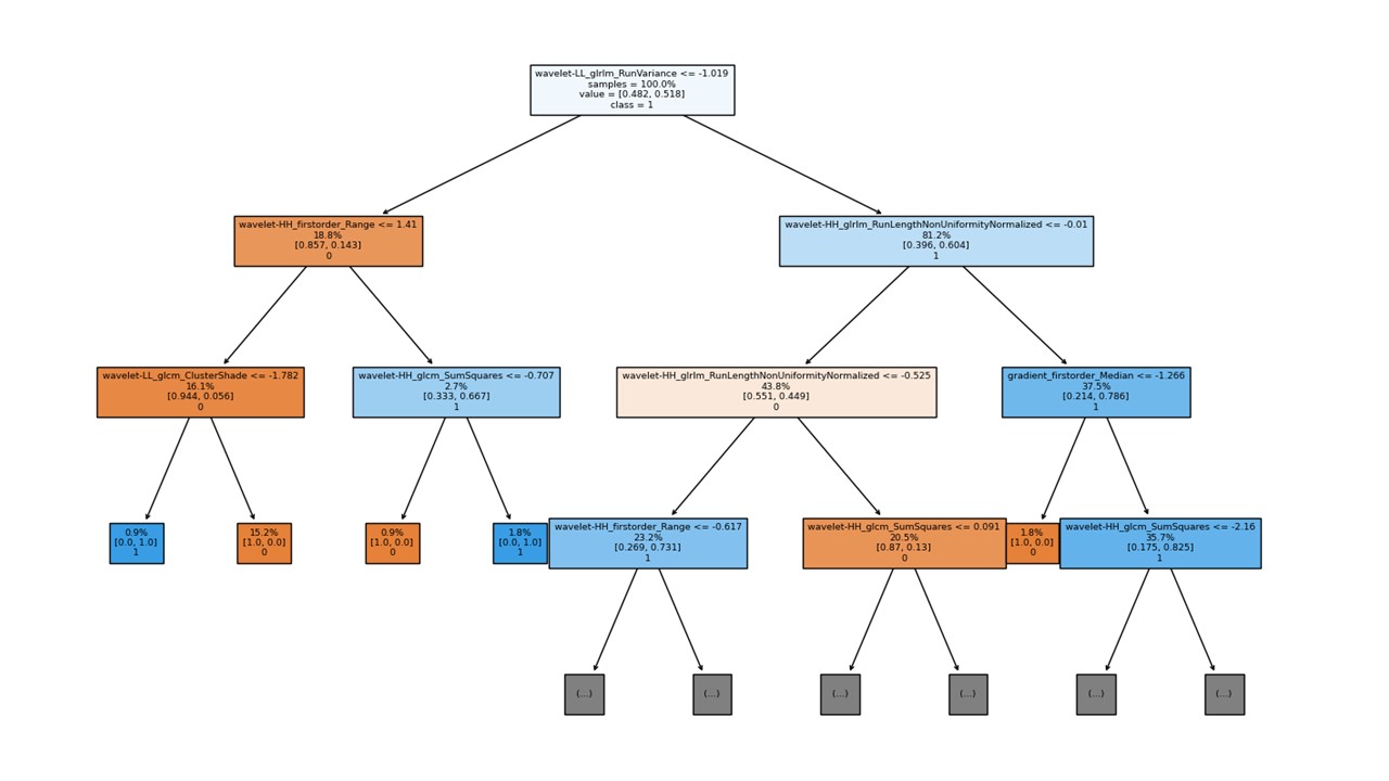
The retrospective study included one hundred and seventy-five CSM patients who underwent surgical treatment. Patients with preoperative conventional magnetic resonance imaging (MRI) were divided into 2 groups according to the changes of the modified Japanese Orthopedic Association (mJOA) scores: the poor outcome group (recovery rate<50%) and the good outcome group (recovery rate≥50%). Segmentation of the narrowest spinal cord was obtained by Spinal Cord Toolbox and manually corrected by experienced radiologists on the axial images. The final data set was randomized into the training set and validation set at the ratio of 7:3. The threshold selection algorithm, univariate feature selection, least absolute shrinkage, and selection operator logistic regression (LASSO), and tree-based feature selection were applied sequentially to select features extracted from MR images. An extremely randomized trees model was constructed. Clinical features were added to the model. Using the area under the receiver operating characteristic curve (AUC), sensitivity, and specificity to assess models’ performance. Further, we attempted to unbox the radiomic feature model. The mean decreases in impurity (MDI), Permutation importance, and SHapley Additive exPlanations (SHAP) were used to measure feature importance. Partial dependence, local dependence, and accumulated dependence were used to analyze the relationship between the outcomes and radiomic predictors. The global surrogate approach was applied to visualize the black-box model.Results
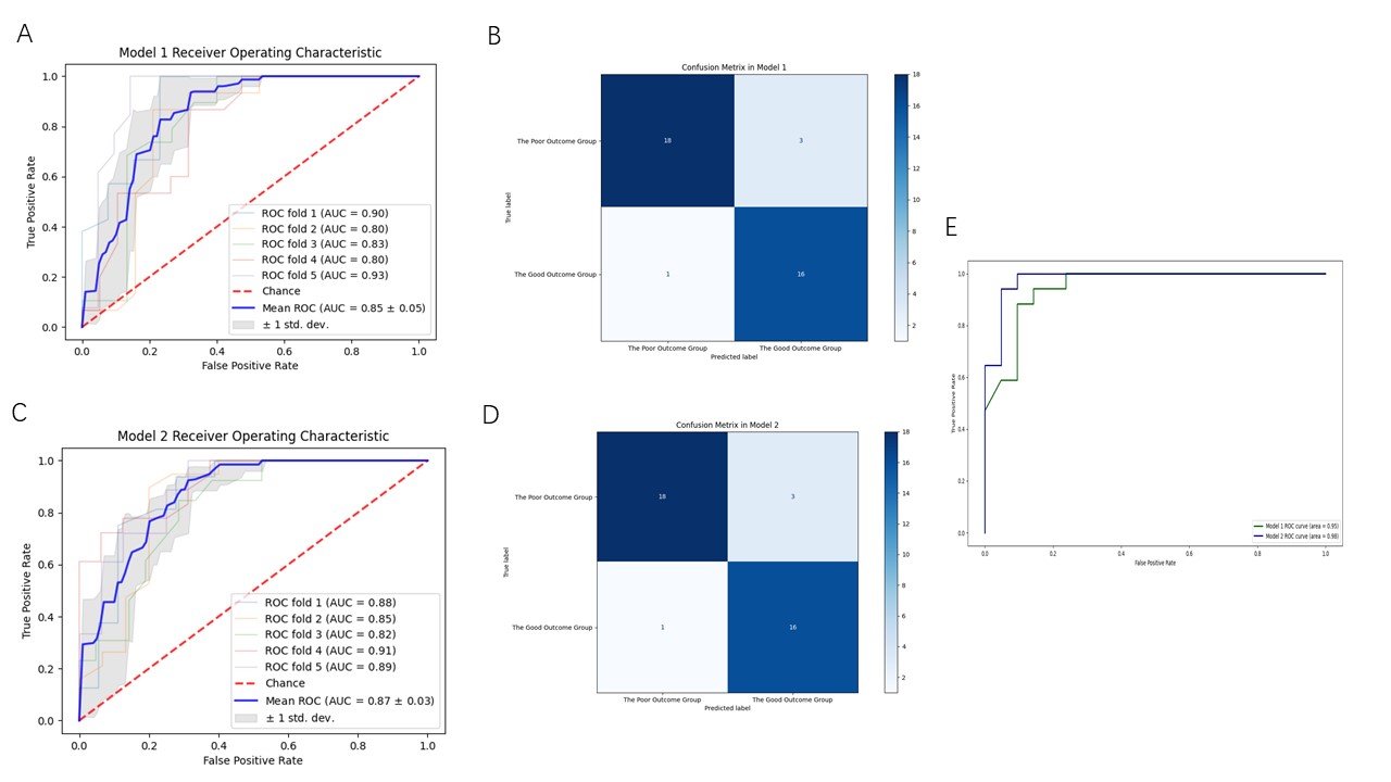
Operation choice (p= 0.000), ages (p= 0.005), anterior-posterior diameter(p=0.035), range of motion(p=0.020) and the MRI-based radiomic signatures were correlated with post-surgical outcomes. The predictive model based on radiomic features and clinical factors performed similarly with the model which was based solely on radiomic features in the training set (AUCs 0.85 vs 0.89, p=0.795) and the validation set (AUCs 0.96 vs 0.97, p=0.795). Run variance, gradient first order median, and cluster shade were related (distance correlation: cluster shade and run variance: 0.494, cluster shade and gradient first order median: 0.393; gradient first order median and run variance: 0.598). No matter which algorithm was closed, the run variance was the most important feature. The non-linear relationships between each feature and outcomes were demonstrated. Along with the increasing of gradient first order median, HH first order range, HH sum squares, LL run variance, and the decreasing of LL cluster shade, patients tended to present a worse outcome. HH run-length non-uniformity normalized played a complicated role in the model. After obtaining a decision tree as a surrogate model (r2=0.928), we found out that run variance was the root node.Conclusions
Radiomics features are potential indicators for predicting CSM patients’ postoperative recovery. Incorporating clinical risk factors and radiomic signatures of MRI images cannot achieve superior postoperative recovery prediction. The run variance was the most important feature in the latter model.Acknowledgements
No acknowledgement found.References
Zhang Peng, Shen Yong, Zhang Ying-Ze, et al. Significance of increased signal intensity on MRI in prognosis after surgical intervention for cervical spondylotic myelopathy.. 2011, 18(8):1080-3.
Yagi Mitsuru, Ninomiya Ken, Kihara Michiya, et al. Long-term surgical outcome and risk factors in patients with cervical myelopathy and a change in signal intensity of intramedullary spinal cord on Magnetic Resonance imaging.. 2010, 12(1):59-65.
Vedantam Aditya, Jonathan Ashish, Rajshekhar Vedantam. Association of magnetic resonance imaging signal changes and outcome prediction after surgery for cervical spondylotic myelopathy.. 2011, 15(6):660-6.
Jun Jae Shin, Byung Ho Jin, Keun Su Kim, et al. Intramedullary high signal intensity and neurological status as prognostic factors in cervical spondylotic myelopathy. 2010, 152(10):1687-1694.
Lindsay A. Tetreault, Joseph R. Dettori, Jefferson R. Wilson, et al. Systematic Review of Magnetic Resonance Imaging Characteristics That Affect Treatment Decision Making and Predict Clinical Outcome in Patients With Cervical Spondylotic Myelopathy. 2013, 38(22S Suppl 1):S89-S110.
Choi Seokmin, Lee Sang-Ho, Lee Ji-Young, et al. Factors affecting prognosis of patients who underwent corpectomy and fusion for treatment of cervical ossification of the posterior longitudinal ligament: analysis of 47 patients.. 2005, 18(4):309-14.
Delphine Zeitoun, Firass El Hajj, Elhadi Sariali, et al. Evaluation of spinal cord compression and hyperintense intramedullary lesions on T2-weighted sequences in patients with cervical spondylotic myelopathy using flexion-extension MRI protocol. 2015, 15(4):668-674.
Daniel W. Apley,Jingyu Zhu. Visualizing the effects of predictor variables in black box supervised learning models[J]. Journal of the Royal Statistical Society: Series B (Statistical Methodology),2020,82(4).
Noam Siegelman,Louisa Bogaerts,Ram Frost. What Determines Visual Statistical Learning Performance? Insights From Information Theory[J]. Cognitive Science,2019,43(12).
Philippe Lambin,Ralph T.H. Leijenaar,Timo M. Deist,Jurgen Peerlings,Evelyn E.C. de Jong,Janita van Timmeren,Sebastian Sanduleanu,Ruben T.H.M. Larue,Aniek J.G. Even,Arthur Jochems,Yvonka van Wijk,Henry Woodruff,Johan van Soest,Tim Lustberg,Erik Roelofs,Wouter van Elmpt,Andre Dekker,Felix M. Mottaghy,Joachim E. Wildberger,Sean Walsh. Radiomics: the bridge between medical imaging and personalized medicine[J]. Nature Reviews Clinical Oncology,2017,14(Suppl.).
van Griethuysen Joost J M,Fedorov Andriy,Parmar Chintan,Hosny Ahmed,Aucoin Nicole,Narayan Vivek,Beets-Tan Regina G H,Fillion-Robin Jean-Christophe,Pieper Steve,Aerts Hugo J W L. Computational Radiomics System to Decode the Radiographic Phenotype.[J]. Cancer research,2017,77(21).
Joshua Bakhsheshian,Vivek A. Mehta,John C. Liu. Current Diagnosis and Management of Cervical Spondylotic Myelopathy[J]. Global Spine Journal,2017,7(6).
Wei Zhang,Hai Yu,Yu-li Zhao,Zhi-liang Zhu. Image encryption based on three-dimensional bit matrix permutation[J]. Signal Processing,2016,118.
Liu Tao,Yang Hui-Lin,Xu Yao-Zeng,Qi Rong-Fu,Guan Hua-Qing. ACDF with the PCB cage-plate system versus laminoplasty for multilevel cervical spondylotic myelopathy.[J]. Journal of spinal disorders & techniques,2011,24(4).Tomosato Yamazaki,Kiyoyuki Yanaka,Kazuya Uemura,Akira Matsumura. Cervical Spondylotic Myelopathy: Surgical Results and Factors Affecting Outcome with Special Reference to Age Differences: In Reply:[J]. Neurosurgery,2003,53(3).
Eiji Wada,Kazuo Yonenobu,Shozo Suzuki,Atsunori Kanazawa,Takahiro Ochi. Can Intramedullary Signal Change on Magnetic Resonance Imaging Predict Surgical Outcome in Cervical Spondylotic Myelopathy?[J]. Spine,1999,24(5).
Sarah H Peckinpaugh. An improved method for computing gray-level cooccurrence matrix based texture measures[J]. CVGIP: Graphical Models and Image Processing,1991,53(6).
Figures