3435
Intravoxel Incoherent Motion Reconstruction with Multi-Orientation Acquisition using Three b-Values1Department of Medical Physics and Biomedical Engineering, Tehran University of Medical Sciences, Tehran, Iran (Islamic Republic of), 2Quantitative MR Imaging and Spectroscopy Group, Research Center for Molecular and Cellular Imaging, Tehran, Iran (Islamic Republic of)
Synopsis
The IVIM model is capable of extracting functional and structural information simultaneously without the injection of contrast agents. The main limitation of this technique is the inaccuracy of the output of this model in low SNR regimes. In this study, we proposed the use of twelve diffusion imaging orientations and three b-values instead of three orthogonal DW imaging and several b-values. Simulation and in-vivo results showed that the proposed method outperforms the conventional IVIM reconstruction method. Improved quality and reproducibility can make this method more practical and attractive in clinical settings.
Introduction
The Intravoxel incoherent motion (IVIM) model is a subfield of diffusion-weighted imaging (DWI) that is based on signal attenuation due to the diffusion phenomenon1,2. In this model, a two-exponential model is proposed to separate diffusion attenuation and perfusion attenuation3. This model has three outputs called diffusion coefficient (D), fraction of blood volume (f), and pseudo-diffusion coefficient (D*), the last two of which are proportional to functional information4. Because this method reveals both structural and functional information, it has seen an increase in recent years for diagnosis in the different fields including liver5, kidney6, breast 7, brain 8, etc. Besides the high potential power of diagnosis, this method does not have high accuracy, especially for perfusion parameters. So far, several studies have been proposed to improve the solution method to increase precision 9-11, and also some studies have been used to optimize the b-values12, 13, but they have not been very effective. So that in most studies, the pseudo-diffusion coefficient which is linked with blood flow4 parameters has not been reported that much.Methods
In this study, we used a minimum of 3 b-values but multi directions DW imaging to improve the accuracy of the output parameters of the images. This study was performed on both computer simulations and in-vivo images. To do in-silico simulation, the tensor of diffusion coefficients and pseudo-diffusion coefficients were selected randomly and of course with the condition of positive semidefinite. A Monte Carlo simulation was performed 100,000 times for SNR 100 and 50. Also, in the in vivo study, images of the brain of a healthy person were performed with a protocol in accordance with the university ethics committee. Conventional IVIM imaging was performed with 11 b-values [0, 50, 100, 150, 200, 300, 400, 500, 600, 700, 800, 1150 $$$\frac{s}{m^2}$$$ ] and 3 orthogonal orientations, then the proposed method imaging was done using 3 b-values [0, 150, 800 $$$\frac{s}{m^2}$$$ ] and 12 orientations.Results
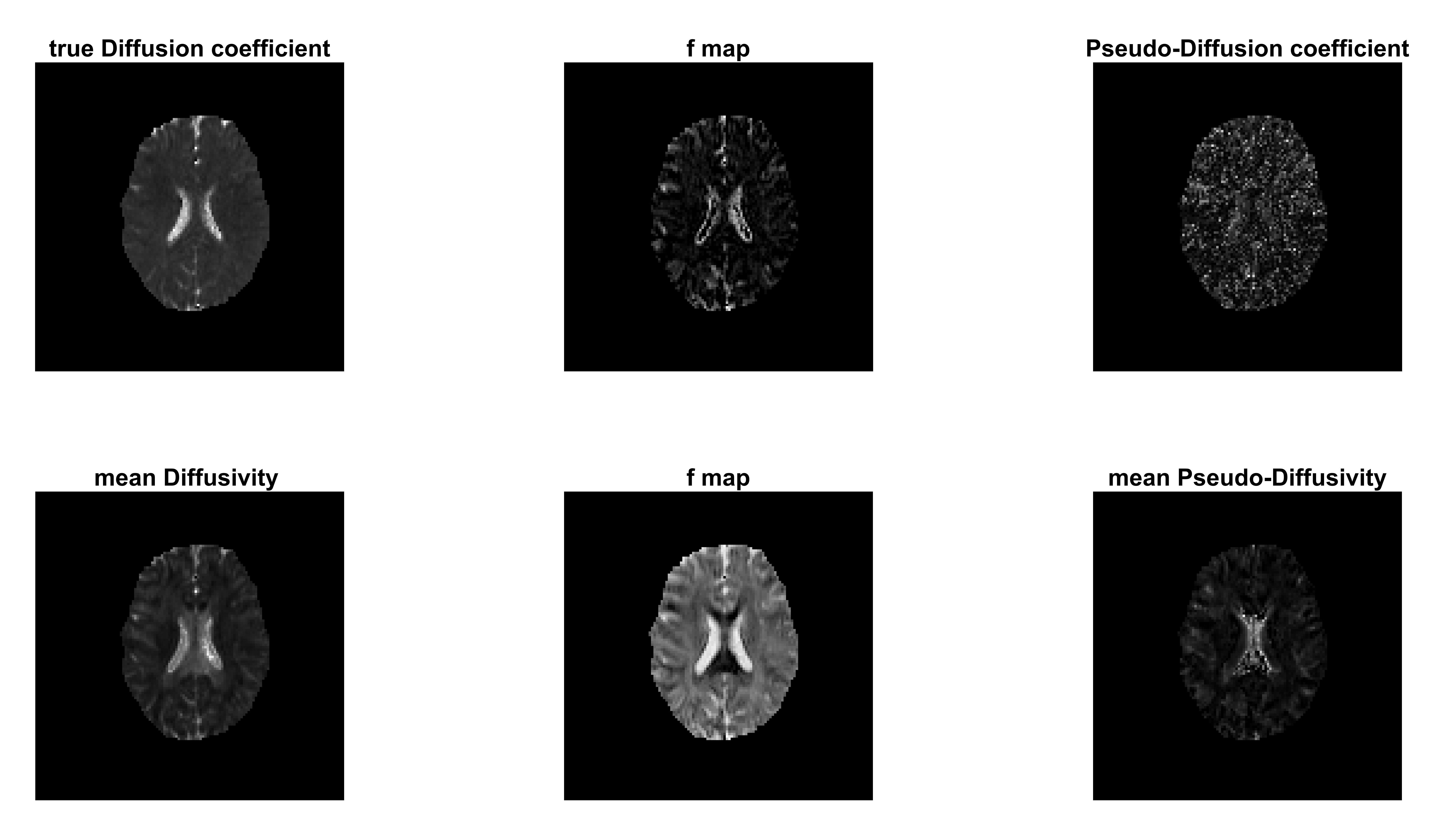
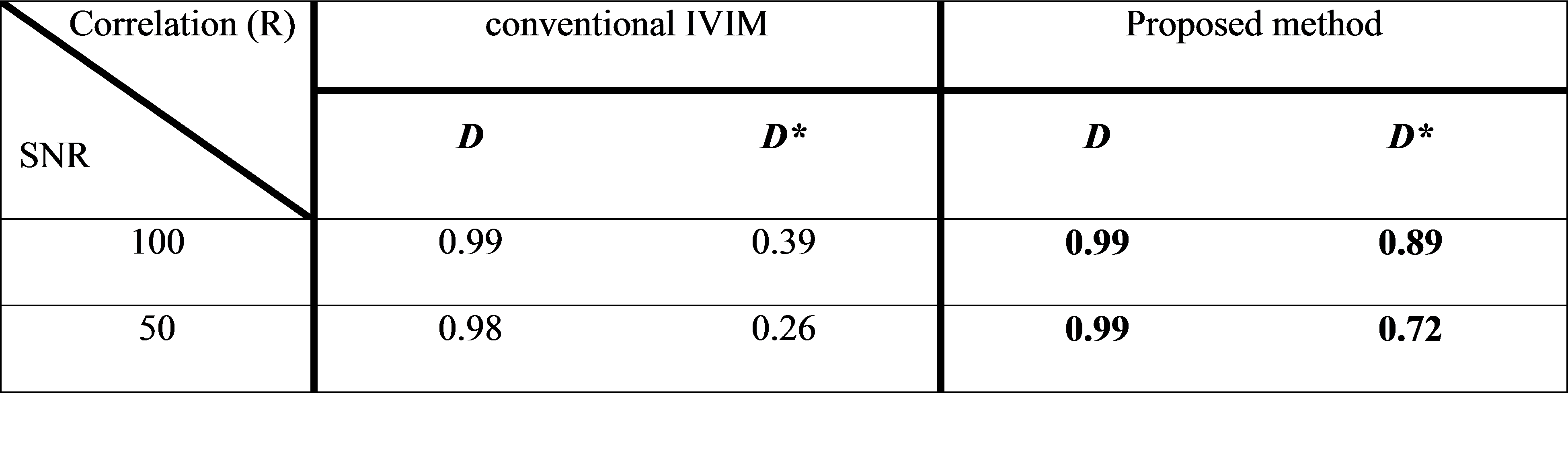
The simulation results clearly show that the output of the parameters of the IVIM model in the proposed method is better than the conventional imaging method. In SNR 100 the results of both methods for the diffusion coefficient are acceptable, although the calculation of the pseudo-diffusion parameter in the proposed method is better than the conventional method. Correlation coefficients of diffusion and pseudo-diffusion coefficient and their estimations in SNR 50 are 0.99 and 0.72 for the proposed method, respectively. In comparison, conventional IVIM imaging correlation coefficients are 0.98 and 0.26 for diffusion and pseudo-diffusion coefficient that shows the proposed method outperforms the conventional method (Table1).Also, the results of in-vivo images showed that especially pseudo-diffusion coefficient maps for the conventional method are almost meaningless and incomprehensible, while the output map of the pseudo-diffusion coefficient in the proposed method is clearly of better quality (Fig1).
Discussion
Theoretically, the IVIM method is very informative because it provides structural and functional information simultaneously and at a suitable resolution. One of the serious limitations in clinics is its inaccuracy, especially for the functional maps; component of the perfusion signal decay is much smaller than the diffusion signal decay, therefore perfusion maps are highly affected in low SNR regime. Our results indicate that multi-direction three b-value DW imaging has robust results in comparison with three-direction multi b-value imaging. Since IVIM imaging is based on measuring the diffusion of water molecules in the direction of motion-sensitive gradients, accurate measurement of parameters depends on the number of DW imaging direction. In the conventional method, only three directions are acquired, therefore measuring IVIM parameters will be only precise if water molecules diffuse either isotropic or just parallel to the gradient directions. However, if the direction of molecular motion is affected by factors such as the presence of nerve fibers in a certain direction, the conventional method will have difficulty in estimating coefficients in even high SNR regimes. Therefore, in complex propagation media such as brain; the more orientations used to measure, the more accurate the parameter estimation will be.Conclusion
IVIM method is data demanding MR imaging technique with functional and structural information estimation that can be used for diagnosis in various organs. Using the proposed method - DW images with 3 b-values and 12 orientations- not only improves the accuracy of the estimated parameters but also more information such as DTI maps can be extracted. IVIM model is a promising method to be used in diagnosis and differentiation of diseases in complex propagation media such as brain.Acknowledgements
No acknowledgement found.References
1. Stejskal, E.O. and J.E. Tanner, Spin diffusion measurements: spin echoes in the presence of a time‐dependent field gradient. The journal of chemical physics, 1965. 42(1): p. 288-292.
2. Le Bihan, D., What can we see with IVIM MRI? Neuroimage, 2019. 187: p. 56-67.
3. Le Bihan, D., et al., Separation of diffusion and perfusion in intravoxel incoherent motion MR imaging. Radiology, 1988. 168(2): p. 497-505.
4. Bihan, D.L. and R. Turner, The capillary network: a link between IVIM and classical perfusion. Magnetic resonance in medicine, 1992. 27(1): p. 171-178.
5. Luciani, A., et al., Liver cirrhosis: intravoxel incoherent motion MR imaging—pilot study. Radiology, 2008. 249(3): p. 891-899.
6. Ichikawa, S., et al., Intravoxel incoherent motion imaging of the kidney: alterations in diffusion and perfusion in patients with renal dysfunction. Magnetic resonance imaging, 2013. 31(3): p. 414-417.
7. Iima, M., et al., Quantitative non-Gaussian diffusion and intravoxel incoherent motion magnetic resonance imaging: differentiation of malignant and benign breast lesions. Investigative radiology, 2015. 50(4): p. 205-211.
8. Federau, C., et al., Cerebral perfusion measurement in brain death with intravoxel incoherent motion imaging. Neurovascular Imaging, 2016. 2(1): p. 9.
9. Fusco, R., M. Sansone, and A. Petrillo, A comparison of fitting algorithms for diffusion-weighted MRI data analysis using an intravoxel incoherent motion model. MAGMA, 2017. 30(2): p. 113-120.
10. While, P.T., A comparative simulation study of bayesian fitting approaches to intravoxel incoherent motion modeling in diffusion‐weighted MRI. Magnetic resonance in medicine, 2017. 78(6): p. 2373-2387. 11. Becker, A.S., et al., Investigation of the pulsatility of cerebrospinal fluid using cardiac-gated Intravoxel Incoherent Motion imaging. Neuroimage, 2018. 169: p. 126-133.
12. Ye, C., et al., Estimation of intravoxel incoherent motion parameters using low b-values. PloS one, 2019. 14(2).
13. Doudou, N.R., et al., Optimization of intravoxel incoherent motion (IVIM): variability of parameters measurements using a reduced distribution of b values for breast tumors analysis. MAGMA, 2020. 33(2): p. 273-281.
Figures