3428
B-value influence on IVIM MRI for juvenile idiopathic arthritis in the knee1Department of Internal Medicine II, Ulm University Medical Center, Ulm, Germany, 2Department of Diagnostic and Interventional Radiology, Ulm University Medical Center, Ulm, Germany
Synopsis
Intravoxel incoherent motion imaging allows the quantification of diffusion and pseudo-perfusion in tissues and can be used as an alternative to conventional T1-weighted scans that require the use of contrast agents. Its benefits for diagnosing juvenile idiopathic arthritis(JIA) in the knee has previously be demonstrated, but usually requires multi-b-value DWI scans with long scan times. In this work we investigate the influence of different b-value combinations on the calculated diffusion and perfusion fraction values and explore the possibility for IVIM MRI of JIA with fewer b-values and shorter scan times.
Introduction
Diffusion-weighted imaging (DWI) is a widely applied quantitative MRI method for the detection of diffusion of water molecules in tissues, however, the calculation of the apparent diffusion coefficient (ADC) with a single-exponential model omits the impact of microperfusion effects on the ADC1. Intravoxel incoherent motion (IVIM) imaging expands the conventional ADC model and provides a valuable tool not only for the calculation of diffusion coefficients but also the quantification of micro-vessel perfusion in tissues from multi-b-value DWI scans2.Patients with juvenile idiopathic arthritis(JIA), one of the most common entities of chronic arthritidis in children and adolescents, are usually examined by use of T1-weighted MR scans with intravenous application of gadolinium-based contrast material for the visualization of inflamed synovial tissue and to assess disease activity and response to therapy. Previous works have shown that perfusion fractions calculated with IVIM MRI improves diagnostic performance and confidence by allowing the visual differentiation of joint effusion and inflamed synovium without the use of contrast agents3.
While multi-b-value DWI scans with longer scan times are necessary to apply the IVIM model and can be challenging when imaging children, it is so far unknown which and how many b-values are required for reliable diffusion and perfusion quantification of JIA in the knee. In this work, we investigated the influence of different b-value combinations and b threshold values on the calculated IVIM parameters in 10 patients with JIA in the knee.
Methods
In this study, 10 patients (mean age: 14 ± 6 years) with known or suspected arthritis of the knee were included. All data were acquired on a 3T whole-body MRI (Skyra, Siemens Medical, Germany). The patients underwent an examination including a survey scan and a readout-segmented multi-shot EPI (RESOLVE) DWI scan with 10 B-values: 0, 50, 100, 150, 200, 300, 400, 600, 800 and 1000mm/s². Further scan parameters were TR/TE = 3400/57ms, resolution of 1.3 x 1.3 x 3mm, 14 slices, FOV of 180 x 180mm and a scan duration of 5:32min during clinical routine.Fitting of the diffusion D, perfusion/pseudo-diffusion D* and perfusion fraction f parameter maps, was performed using a bi-exponential fitting model:
$$S = S_0( (1-f) exp(-bD) + f exp(bD*) )$$
with the measured signal intensity without diffusion-weighting (b=0 s/mm²) used as $$$S_0$$$ .
Parameter fitting was performed with the commonly used “segmented” two-step fit: first, D and f were calculated from images with b-values above a certain threshold b-value (here called b-threshold) where only negligible influence of the pseudo-diffusion was expected, simplifying the fitting model to a mono-exponential signal decay function. Subsequently, these D and f values were used for the perfusion coefficient calculation in the bi-exponential model.
Three ROIs were manually drawn in muscle, synovial membrane and effusion for each patient data set from which the mean diffusion coefficient and perfusion fraction was calculated.
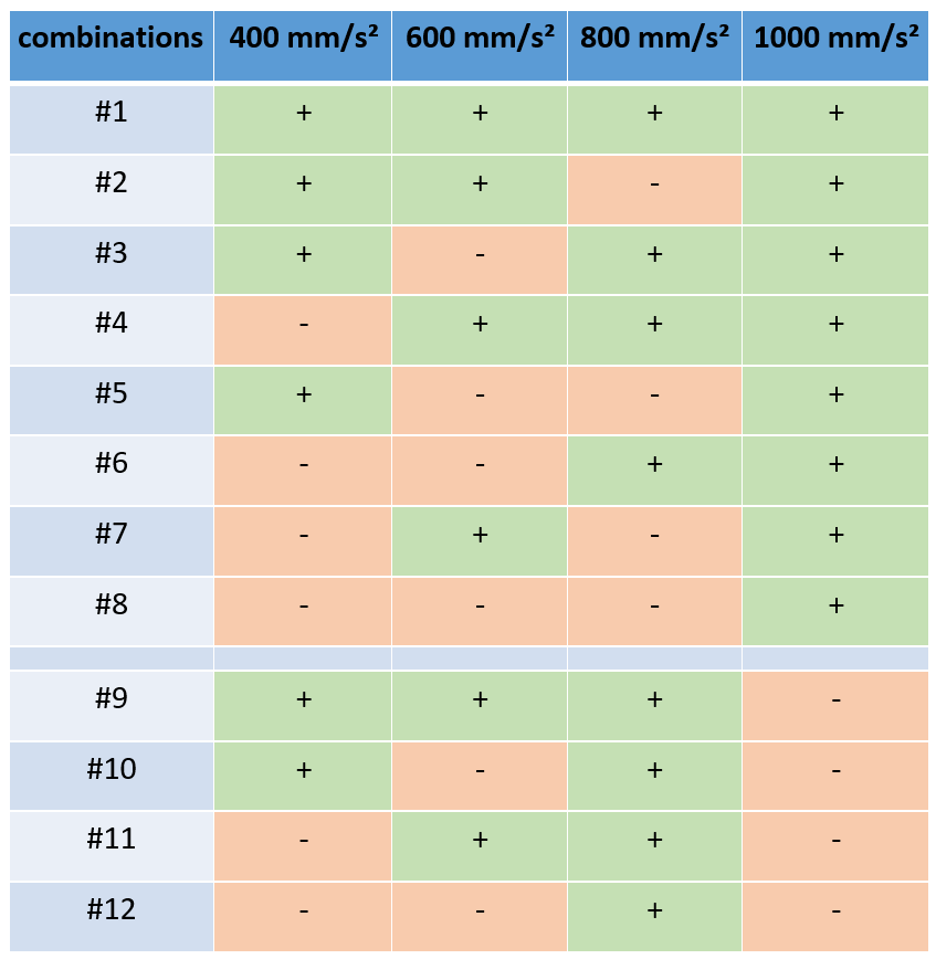
The IVIM parameters were calculated for 12 b-value combinations (see table 1), with the four smallest b-values (50, 100, 150, 200) being included in all combinations, and four b-threshold values (50, 100, 150 and 200mm/s²). D- and f-values calculated from combination #1 (using all measured b-values) acted as reference values to which all other calculated D- and f-values were compared to and the relative difference was calculated (for b-threshold values 50, 100 and 150mm/s²).
Results
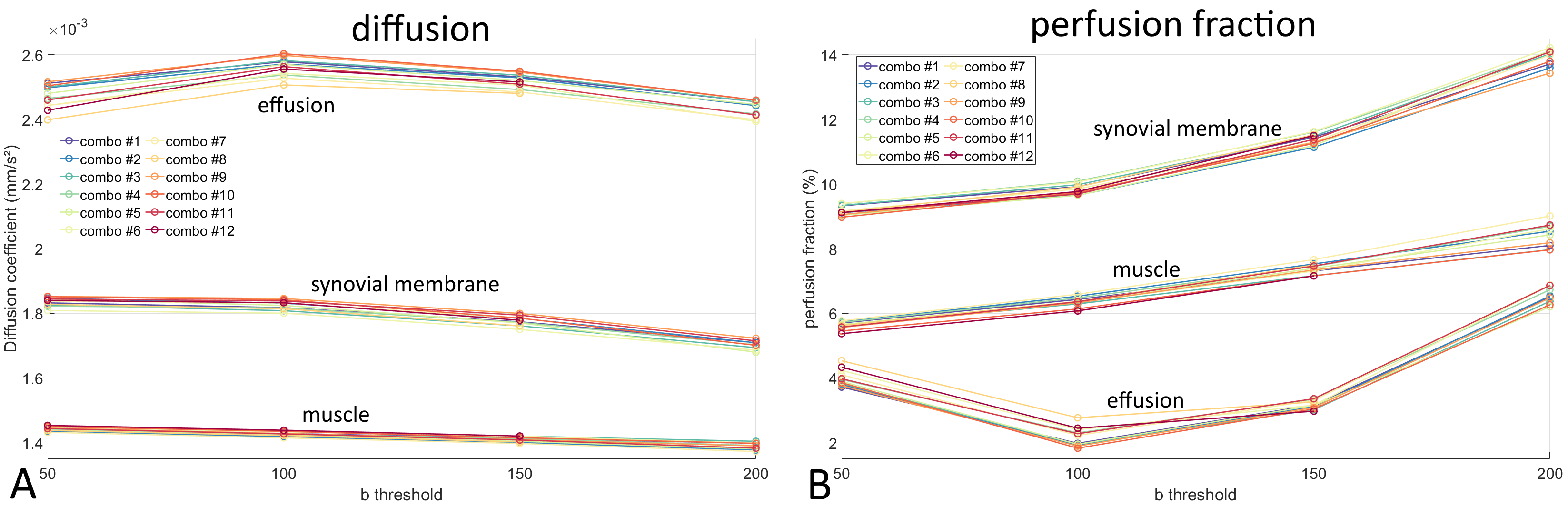
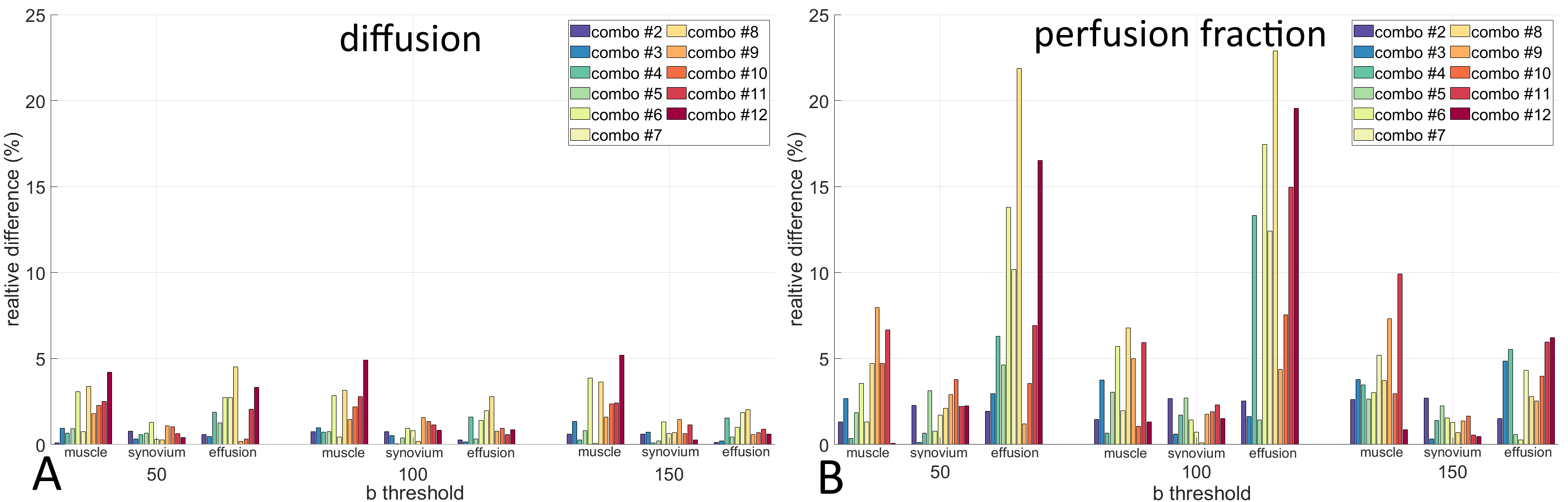
Overall, the calculated D and f values were comparable to those of previous publications [Hilbert Neubauer]. Figure 1 shows the calculated diffusion (A) and perfusion fraction (B) values for muscle, synovial membrane and effusion for all 12 b-value combinations in dependence of the b-threshold value. While no clear influence of the b-threshold could be observed on the D values, f distinctly increased with a larger b-threshold.Relative differences for calculated diffusion (A) and perfusion fraction (B) values between combination #1 and all other b-value combinations are displayed in Figure 2. While the choice of b-values clearly has a large influence on the perfusion fraction values in effusions, with b combinations omitting smaller b-values showing the largest differences of up to 22.6%, only small differences between b combinations (rarely over 5%) could be observed for muscle and synovium f values and D coefficients in general (largest difference 5.2%). For perfusion fraction values a b-threshold of 150mm/s² led to smaller differences between b combinations than the lower threshold values of 50 and 100mm/s².
Discussion and conclusion
Overall it could be demonstrated that IVIM parameters could be reliably calculated even with b-value combinations comprising only few b-values. Since diffusion coefficient calculation is mostly influenced by the highest measured b-value, the negligible difference in D values between the b combinations was to be expected.With the IVIM effect usually being small, low b-values are essential for providing reliable results for the calculated pseudo-diffusion and perfusion fraction. Thus, b combinations with fewer b-values in the intermediate range resulted in a larger variation of calculated f-values, especially visible in tissue with low perfusion such as effusions. Additionally, usage of higher b-thresholds led to smaller differences in perfusion fraction values between b combinations.
In conclusion, IVIM MRI for JIA in the knee can be reliably performed with as few as 5 b-values, allowing for shorter scan times suitable for examinations of children and juveniles.
Acknowledgements
The authors thank the Ulm University Center for Translational Imaging MoMAN for its support.References
[1] Le Bihan D. et. al. MR imaging of intravoxel incoherent motions: application to diffusion and perfusion in neurologic disorders. Radiology (1986) 161 (2): 401–7.
[2] Sigmund E.et al. Intravoxel incoherent Motion (IVIM) imaging of tumor microenvironment in Locally Advanced Breat Cancer: Magn: Reson. Med. 2011 65(5):1437-1447
[3] Hilbert F. et.al. Intravoxel incoherent motion magnetic resonance imaging of the knee joint in children with juvenile idiopathic arthritis. Pediatr. Radiol. (2017) 47:681-690
Figures