3426
Relevance of NODDI to Characterise In Vivo the Microstructural Abnormalities of Multiple Sclerosis Cortex and Cortical Lesions: A 3T Study
Elisabetta Pagani1, Paolo Preziosa1,2, Raffaello Bonacchi1,2, Laura Cacciaguerra1,2,3, Massimo Filippi1,2,3,4,5, and Maria A. Rocca1,2,3
1Neuroimaging Research Unit, Division of Neuroscience, IRCCS San Raffaele Scientific Institute, Milan, Italy, 2Neurology Unit, IRCCS San Raffaele Scientific Institute, Milan, Italy, 3Vita-Salute San Raffaele Unversity, Milan, Italy, 4Neurorehabilitation Unit, IRCCS San Raffaele Scientific Institute, Milan, Italy, 5Neurophysiology Service, IRCCS San Raffaele Scientific Institute, Milan, Italy
1Neuroimaging Research Unit, Division of Neuroscience, IRCCS San Raffaele Scientific Institute, Milan, Italy, 2Neurology Unit, IRCCS San Raffaele Scientific Institute, Milan, Italy, 3Vita-Salute San Raffaele Unversity, Milan, Italy, 4Neurorehabilitation Unit, IRCCS San Raffaele Scientific Institute, Milan, Italy, 5Neurophysiology Service, IRCCS San Raffaele Scientific Institute, Milan, Italy
Synopsis
In multiple sclerosis (MS), cortical damage is a relevant predictor of clinical disability. We applied Neurite orientation dispersion and density imaging (NODDI) to characterize microstructure of normal-appearing cortex (NA-cortex) and cortical lesions (CLs) and their relations with disease clinical phenotypes and disability. We found that a significant neurite loss occurs in MS NA-cortex, being more severe with longer disease duration, more severe disability and progressive MS. CLs show a further reduction of neurite density, together with an increased extracellular space, possibly due to inflammation and gliosis, and a reduced dispersion suggestive of increased tissue coherence and simplification of neurite complexity.
Introduction
In multiple sclerosis (MS), cortical damage is a relevant predictor of clinical disability, but MRI measures more specific to cortical pathology are needed. Neurite orientation dispersion and density imaging (NODDI) model1 is a multi-compartment diffusion model to better evaluate the complexity of brain microarchitecture. The aim of this study was to characterize, using NODDI, the microstructural abnormalities of normal-appearing cortex (NA-cortex) and cortical lesions (CLs) and their relations with disease phenotypes and clinical disability in a relatively large cohort of MS patients.Methods
Brain 3D T1-weighted, FLAIR, double inversion recovery (DIR) and diffusion-weighted (DW) sequences were acquired from 172 MS patients (101 relapsing-remitting [RR], 71 progressive [P] MS) and 62 healthy controls (HC). The cortex was segmented from 3D T1-weighted sequence and CLs were quantified on DIR. CLs and NA-cortex masks were then transformed into DW space (Figure 1). Using NODDI, intracellular volume fraction (ICV_f), representing neurite volume, extracellular volume fraction (ECV_f) and orientation dispersion index (ODI), reflecting neurite orientation variability, were assessed in NA-cortex and CLs. Between-group comparisons and correlations with clinical and structural MRI measures were investigated.Results
One hundred and seventeen (68%) MS patients had ≥1 CL. MS NA-cortex had a significant lower ICV_f vs HC NA-cortex (p<0.001). CLs showed a significant increased ECV_f (p<0.001) and decreased ICV_f and ODI compared to NA-cortex of HC (p<0.001) and MS (p=0.006 and <0.001) (Figure 2). Compared to RRMS, PMS had a significant decreased NA-cortex ICV_f (p=0.03) and ODI (p=0.02). Higher burden of CLs (p<0.001) was found in PMS vs RRMS, without microstructural differences. In MS patients, NA-cortex ICV_f, ECV_f and ODI were significantly correlated with disease duration, EDSS, white matter lesion volumes, CL volumes and whole brain and gray matter atrophy (r from -0.51 to 0.71, p from <0.001 to 0.02).Discussion
A significant neurite loss occurs in MS NA-cortex, being more severe with longer disease duration, more severe disability and PMS. CLs show a further reduction of neurite density, together with an increased extracellular space, possibly due to inflammation and gliosis, and a reduced ODI suggestive of increased tissue coherence and simplification of neurite complexity.Conclusions
NODDI is reliable and clinically relevant to investigate in vivo the heterogeneous pathological processes affecting MS cortex.Acknowledgements
Funding. This study is supported by a senior research fellowship from FISM – Fondazione Italiana Sclerosi Multipla – cod. 2019/BS/009 and financed or co-financed with the ‘5 per mille’ public funding.References
1. Zhang H, Schneider T, Wheeler-Kingshott CA, Alexander DC. NODDI: Practical in vivo neurite orientation dispersion and density imaging of the human brain. NeuroImage 2012; 61:1000–1016.Figures
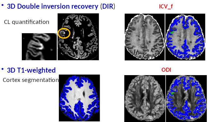
Figure 1. The postprocessing is
shown for a representative MS patient: after the segmentation of cortical
lesions and gray matter, masks are overlapped on the maps of intracellular
volume fraction (ICV_f) and orientation dispersion (ODI).
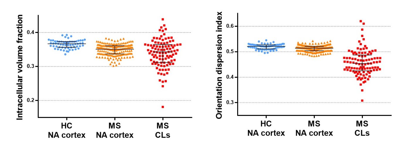
Figure 2. Scatter plots of NODDI
indexes obtained within the normal-appearing cortex (NA-cortex) and cortical
lesions (CLs) of MS patients and healthy controls.