3420
A New Phantom to Study a Restricted Diffusion Introduction
Sergey Magnitsky1
1CHOP, Philadelphia, PA, United States
1CHOP, Philadelphia, PA, United States
Synopsis
Diffusion-weighted imaging is instrumental in the evaluation of bone quality. However, an interpretation of the data obtained from porous material is complex due to the effects of restricted diffusion. In this study, we are presenting a new restriction diffusion phantom, which was developed for an optimization of acquisition protocols for bone studies. The phantom consists of microscopic-slides separated by glass spheres (~10 μm). The space between slides was filled with water. NMR data were collected and diffusion-properties of the phantom were documented. The proposed phantom can be easily replicated in any laboratory and will assist in investigations of restriction diffusion.
Introduction
Different patterns of restricted diffusion were detected in various bone conditions. To model restricted diffusion, investigators used phantoms made of natural products like celery stems. These phantoms are not stable and not reproducible. No commercially available restricted diffusion phantoms are available at present. The goal of this study was the development of a simple restricted diffusion phantom to detect:1) the effect of the impermeable boundary on diffusion data, 2) determine b/q-values when the free and restricted water becomes different, 3) probe angular properties of restricted diffusion. This phantom will be utilized for an optimization of acquisition parameters of MRI experiments to study restricted diffusion of water and fat in bones.Methods
Hollow glass spheres with a diameter of 9-13 μm (Sigma-Aldrich) were spread with a painting brush evenly on a surface of 10 microscope glass slides (75 x 38 mm, Fisher Scientific). The slides were put on each other and tighten together with a string. The stuck was immersed in a container with tap water. After 60 min, the phantom was removed from the water and wax was applied on the edges of the phantom to prevent drying. Experiments were conducted at 4.7 T horizontal bore magnet interfaced with Varian console. A two cm surface transmit-received coil was utilized for the acquisition of NMR data. The phantom was placed into the magnet and the diffusion-weighted stimulated echo experiment was performed with the following parameters: TR=10 s, TE = 10 ms, diffusion gradient was varied 0-24 G/cm, the angel of the diffusion gradient was varied 0-900, d = 20 ms, D = 450 ms.Results
Figure 1 shows the normalized q-plot (q=(1/2p)gGd) of the NMR signal intensity of free water and water in the phantom. The Green symbols depict free water diffusion. The Blue symbols present the water signals in the phantom with the diffusion gradient applied parallel to the surface of the glass slides. The Red symbols show water diffusion in the phantom with the diffusion gradient applied perpendicular to the surface of the glass slides. The NMR signal intensity of water molecules inside of the glass phantom is following the free diffusion pattern only in the parallel orientation of the diffusion gradient and the surface of glass slides. Rotation of the diffusion gradient by 90-degree to the surface of the slides revealed the significant deviation from the free diffusion pattern and the presence of local minimums (Figure 1 red symbols). These phenomena were described by Callaghan [1] and reflected the presence of impermeable boundaries. In our experiments, the minimums were detected at q(min1) = 70534 and q(min2) = 160305 1/m. A calculation of characteristic dimension based on this q-values yields a distance of 13.3 and 12.3 mm, which are in a good agreement with dimensions of the spheres used in our phantom as spacers. The orientation of diffusion boundaries in the human bones has a convoluted orientation. To simulate this situation, we conducted a series of experiments with different orientations of the diffusion gradient. Figure 2 shows the q-plots of the NMR signal intensity at different angles between the diffusion gradient and the surfaces of the glass slides. The minimums, typical for the restricted-diffusion, gradually disappeared from the curves, and the shape of the line transforms into the free diffusion pattern with the decrease of the diffusion gradient angle.Discussion and conclusions
In this study, we developed a phantom to simulate diffusion data in porous compartments of human bone. This simple phantom with known geometry allowed us: 1) to detect effects of the impermeable boundary on diffusion data, 2) determine parameters of the diffusion experiments when the free and restricted water exhibit different behavior, 3) probe angular properties of restricted diffusion. Characterization of bone porosity plays an important role in the diagnosis of bone degenerative disorders. The developed phantom permitted us to model the restricted diffusion in human bones and measure the characteristic dimension of the compartment. The red curve on Figure 1 is an example of restricted diffusion, while the green and blue symbols are illustrations of free diffusion. A significant deviation of free and restricted diffusion curves was detected at q value more than 25000. The calculation of the compartment dimensions based on water diffusivity (D = 2.3 x10⁻⁵cm2/sec) and the theory developed by Callaghan revealed ~12.8 μm, which is in good agreement with the design of our phantom. A variation of the orientation of the diffusion gradient allowed us to test the effect of un-parallel boundaries on the diffusion pattern. The proposed phantom can be easily replicated in any lab and further modified to mimic any size of restriction compartments since the hollow spheres of different sizes are commercially available. We built several copies of the phantom and the measurements of the distance in different copies were the same with <5% variation. The similar results obtained from a different copy indicate about good reproducibility of the proposed phantomAcknowledgements
I would like to thank Dr. S Pickup from the University of Pennsylvania for the help with MRI acquisition and useful discussions.References
1. Callaghan, P.T., NMR imaging, NMR diffraction and applications of pulsed gradient spin echoes in porous media. Magnetic Resonance Imaging, 1996. 14(7-8): p. 701-9.Figures
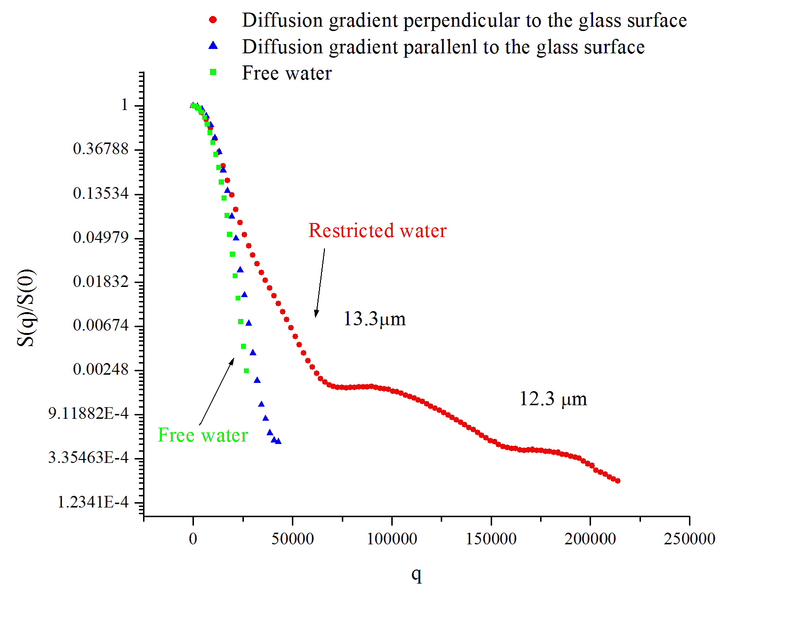
Figure 1 Normalized NMR signal intensity of free (green, blue) and restricted
(red) water molecules at different orientation of the diffusion gradient
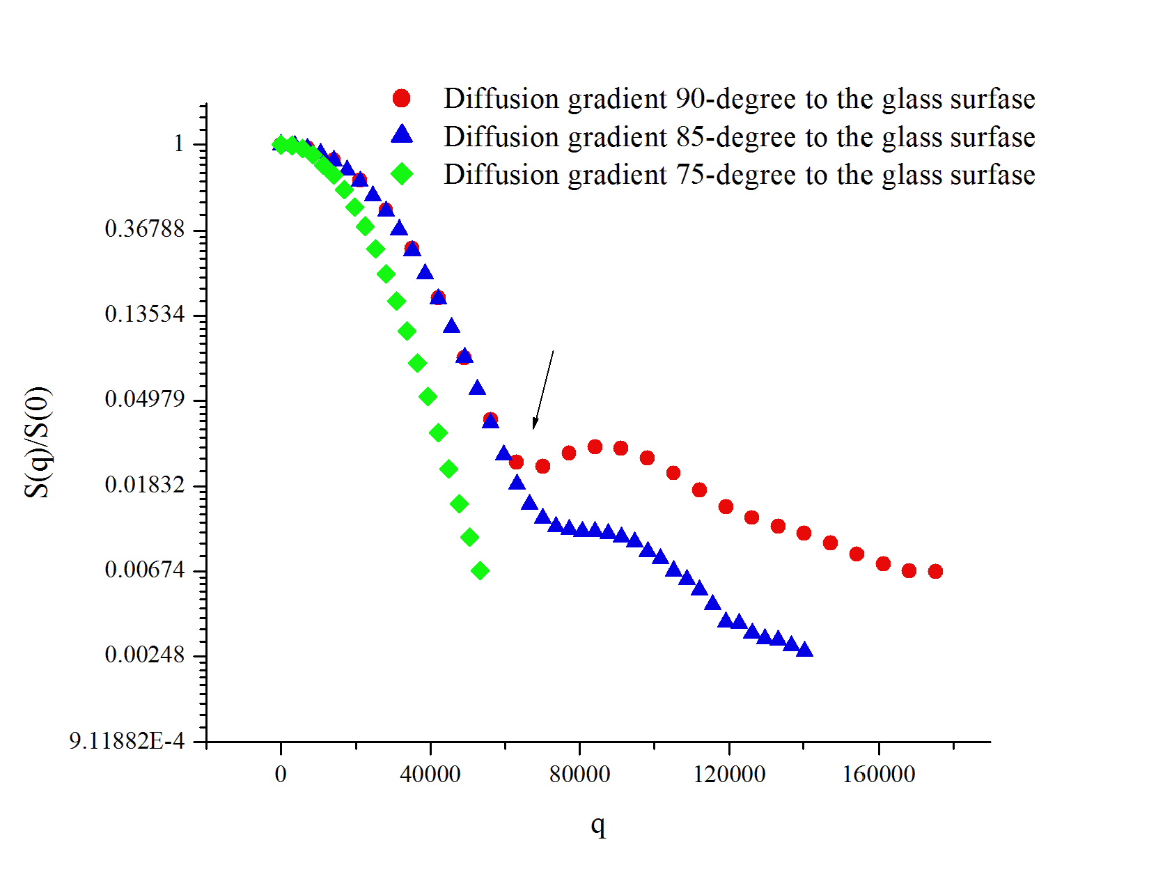
Figure 2 Normalized NMR signal intensity of restricted water molecules at
different orientation of diffusion gradient