3412
Measurement of intraventricular temperature in the whole brain using second order motion compensation DTI1Tokai university hospital, Kanagawa, Japan, 2Tokai University School of Medicine, Isehara, Japan, 3Kanazawa University, Kanazawa, Japan, 4Tokai university hospital, Isehara, Japan, 5Philips Japan, Tokyo, Japan, 6RIKEN Center for Brain Science, saitama, Japan, 7Course of Electrical and Electronic Engineering, Graduate School of Engineering, Tokai University, Isehara, Japan
Synopsis
The intraventricular cerebrospinal fluid (CSF) temperature calculated from the diffusion coefficient is affected by the CSF pulsation. Therefore, we investigated the second-order motion compensation DTI (2nd-MC DTI) in consideration of fractional anisotropy (FA) for the CSF to the determination of the intraventricular temperature to improve that accuracy. The measurement of the intraventricular temperature with 2nd-MC DTI showed the least SD and can be more accurately estimated than conventional DTI.
INTRODUCTION
Intraventricular temperature can be estimated by measuring the diffusion coefficient in the ventricular CSF 1. However, there is a drawback that the CSF pulsation and flow cause an error in the estimation of the intraventricular temperature. To overcome this limitation, we come up with using second order motion compensation (2nd-MC) diffusion tensor imaging (DTI) for intraventricular temperature measurement. On the other hand, the difference of the diffusion coefficient is known in the ventricles among each motion-proving-gradient (MPG) direction in DTI 2. Thus, we propose the calculation of intraventricular temperature considering the fractional anisotropy (FA) of the CSF. The purpose of this study was to investigate whether applying the 2nd-MC DTI with FA processing improves the determination of the intraventricular temperature, compared with conventional-DTI (c-DTI).METHODS
The temperature dependence of FA was investigated to determine the threshold of FA processing. The phantom was scanned with a 3.0 T MR system (Ingenia R5.6; Philips Healthcare, Best, the Netherlands). A bottle as the phantom was filled with artificial CSF (R&D SYSTEMS). The phantom was heated to 40 degrees C in a thermostatic water bath (NTT-20S, Eyela), and then cooled down to approximately 30 degrees C. During the cooling process the sample temperature was monitored by fluorescent fiberoptic thermometer while scanning with 2nd-MC DTI. Ten subjects were examined at a 3.0 T MR system. We evaluated the accuracy of the measurement of the intraventricular temperature between c-DTI and 2nd-MC DTI (Fig.1). Each DTI of whole brain was obtained. We performed two-dimensional single-shot echo-planar imaging (EPI) DTI with the following parameters: TR, 8000 ms; TE, 119 ms; flip angle, 90 degrees; field of view, 220 × 220 mm2; matrix,128 × 128; slice thickness, 4.0 mm; number of slices, 30; EPI factor, 51; diffusion-encoding directions, 15; sensitivity encoding factor, 3; and scan time, 2 min 24 sec. To measure the diffusion coefficient of the CSF, the optimal b value (i.e., 400 s/mm2) based on the literature 3,4 was used. Diffusion coefficient was calculated from the signal intensity of DTI in the all ventricles, and then FA map was analyzed. Since CSF was considered to be free water isotropic diffusion, the calculated pixel values were excluded from the intraventricular temperature map if the FA value exceeded the threshold determined from the phantom study. After FA processing, intraventricular temperature map was generated from diffusion coefficient. The differences for the intraventricular temperature and standard division (SD) in the whole ventricles were assessed using Friedman test and a post-hoc test among each method, i.e., (1)c-DTI, (2)c-DTI with FA processing, (3) 2nd-MC DTI, (4) 2nd-MC DTI with FA processing.RESULTS and DISCUSSION
In the phantom study, FA of the artificial CSF and distilled water were not affected by temperature variation. Since no FA larger than 0.1 were measured, the threshold for the FA processing was determined to be 0.1. Examples of intraventricular temperature maps on whole brain in two methods with FA processing are shown in Fig.2. The intraventricular maps of 2nd-MC DTI with FA processing showed higher uniformity. While some pixels around the foramen of Monro have been removed by FA processing, the intraventricular temperature map of c-DTI with FA processing still showed un-uniformity . The Friedman tests demonstrated significant differences for both the intraventricular temperature and SD in the whole ventricles (Figs.3, 4). The intraventricular temperatures for both DTI with FA processing significantly lower than those without FA processing on the post-hoc test. This result suggests that FA processing allowed the correction for overestimation of diffusion coefficient due to CSF pulsation. The 2nd-MC DTI with FA processing had a significantly smaller SD compared with c-DTI and c-DTI with FA processing on the post-hoc test. Small SD estimation is of great importance as a robust measurement. Since the third ventricle is known to have a to-and-fro motion5, the CSF flow is expected to have not only simple laminar flow but also turbulence. The temperature elimination in turbulent flow is impossible with the use of 2nd-MC DTI alone. Combining 2nd-MC DTI and FA processing is assumed to be an optimal method to improve accuracy.CONCLUSION
The 2nd-MC DTI with FA processing may improve the accuracy to determine the intraventricular temperature in whole brain, compared with c-DTI.Acknowledgements
No acknowledgement found.References
1. Delannoy J, et al. Magn Reson Med. 1991;19:333-9. 2. Kozak LR, et al. Acta Paediatr. 2010;99(2):237-43. 3. Saritas EU, et al. IEEE Trans Med Imaging. 2011; 30:424-37. 4. Tofts PS, et al. Magn Reson Med. 2008;59:190-5. 5. Matsumae, et al. J Neurosurg. 2014;120(1):218-27.Figures
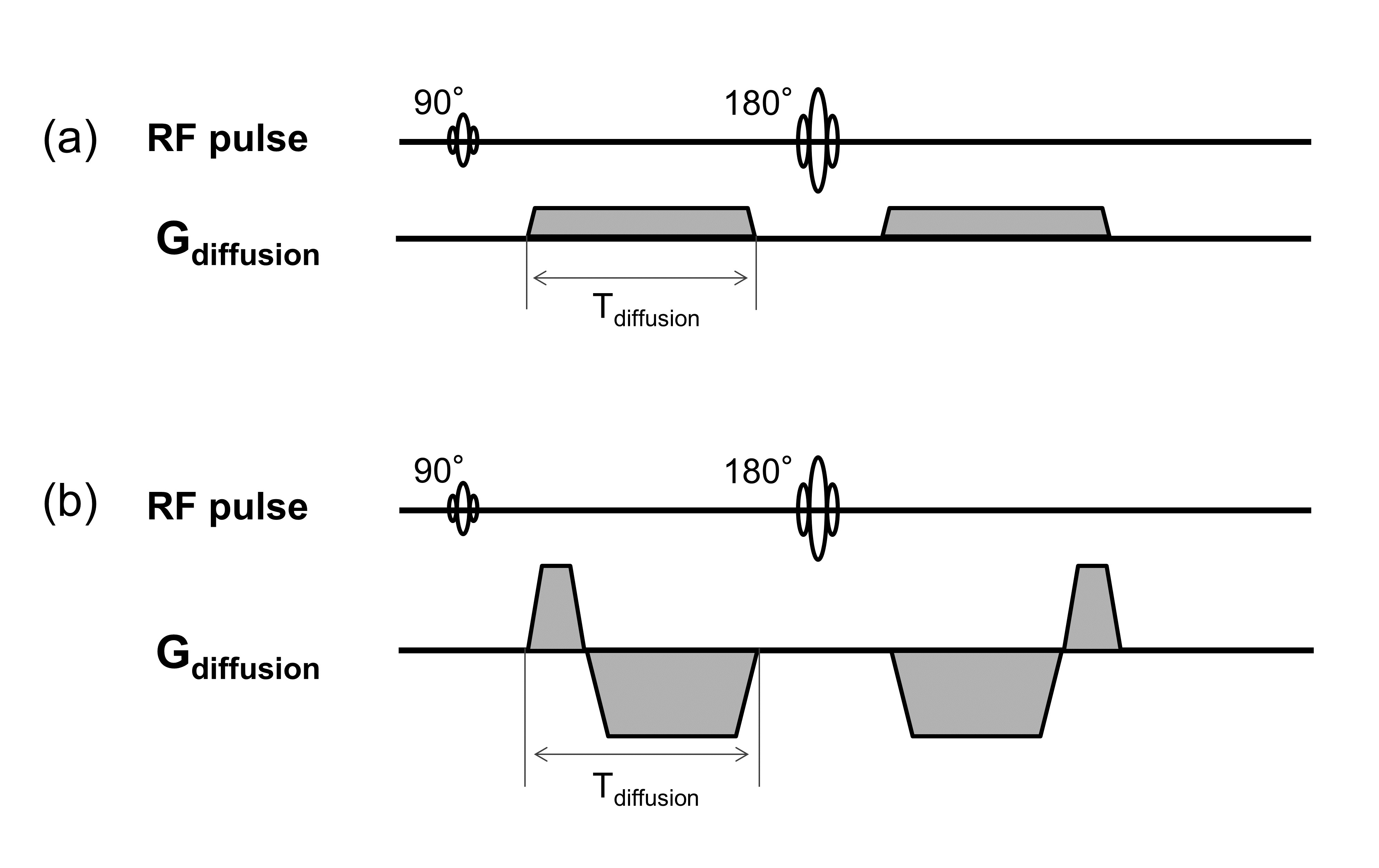
Figure1
The second order motion compensation (2nd-MC) can be achieved by using suitable second moment nulling gradient waveforms. (a) conventional DTI and (b) 2nd-MC DTI.
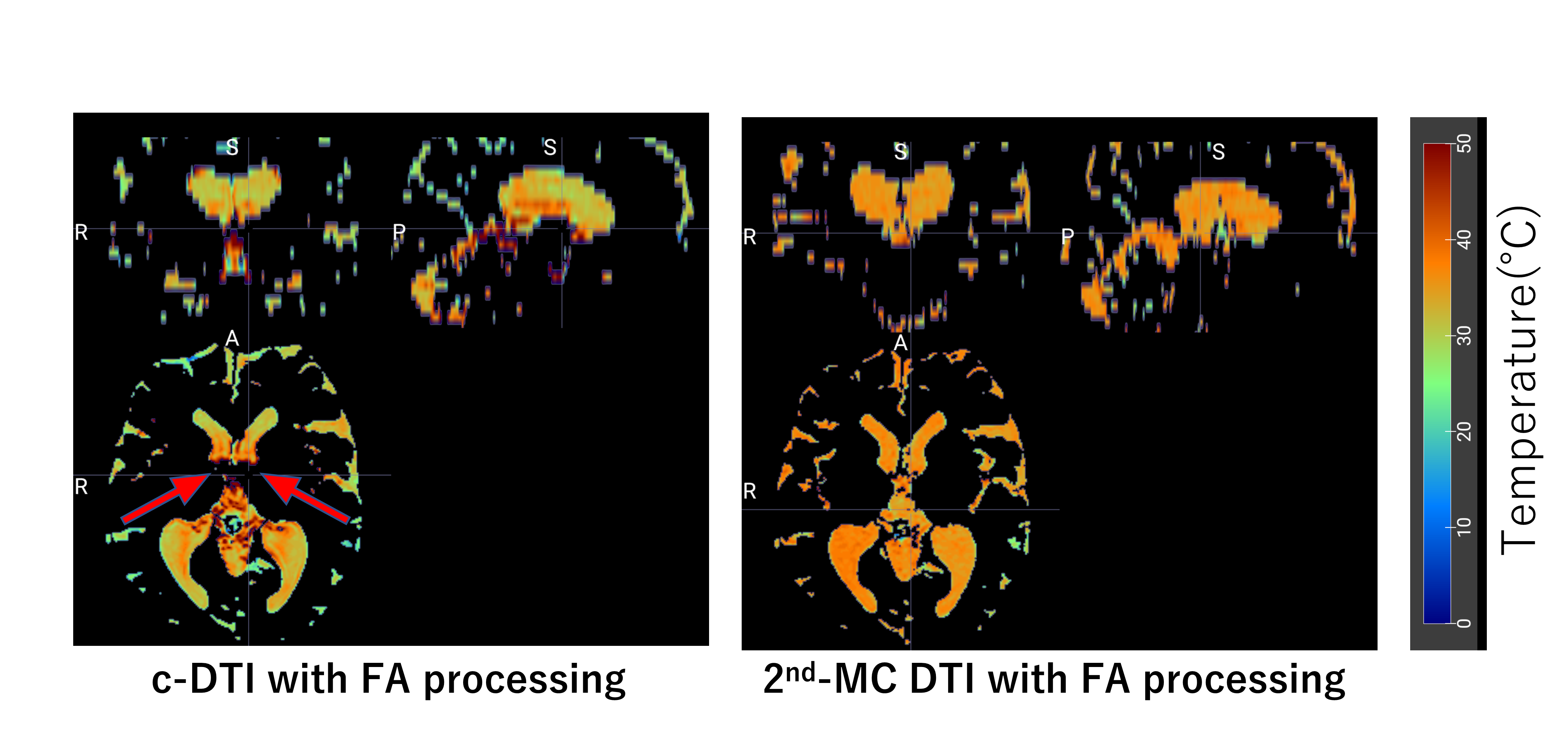
Figure 2
Examples of intraventricular temperature maps for the two DTI techniques with fractional anisotropy (FA) processing. The red arrows indicate some pixels around the foramen of Monro removed by FA processing.
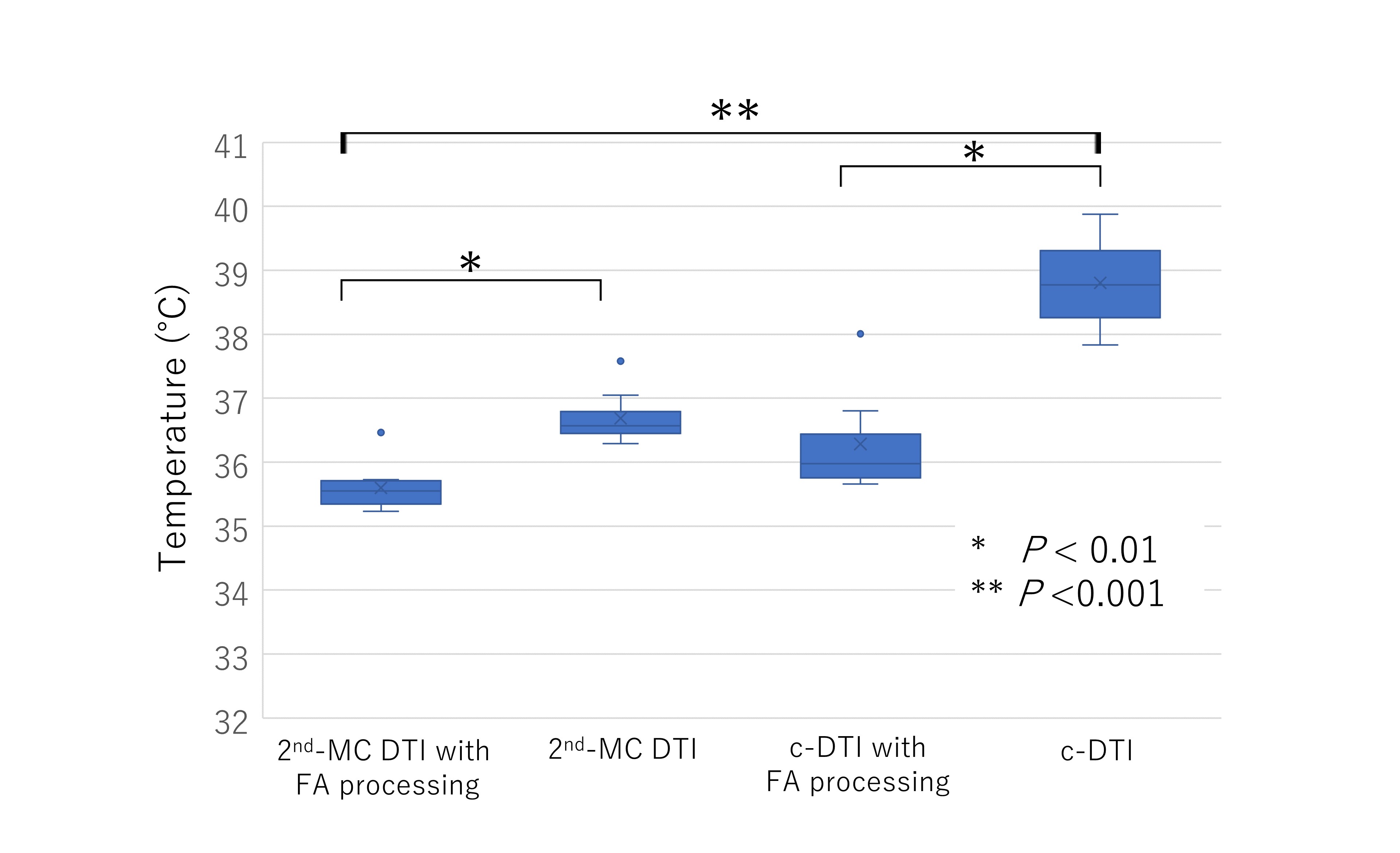
Figure 3
Comparison of intraventricular temperature among four DTI methods in whole ventricles. Asterisk indicates P < 0.01; double asterisk, P < 0.001.
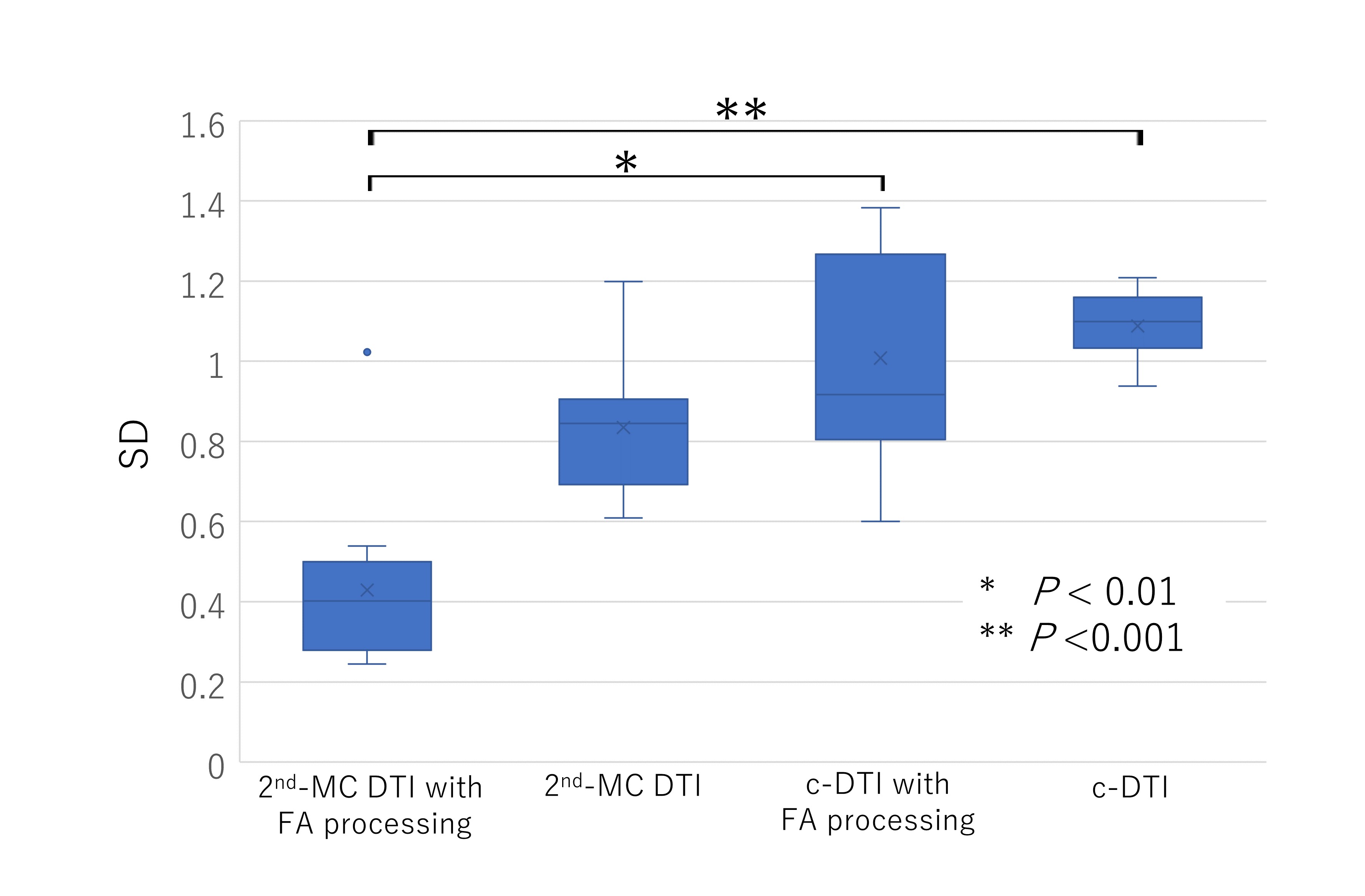
Figure 4
Comparison of standard division (SD) among four DTI methods in whole brain. Asterisk indicates P < 0.01; double asterisk, P < 0.001.