3389
Intraoperative arterial spin labeling (iASL) reliably depicts functional networks during neurosurgery
Thomas Lindner1, Hajrullah Ahmeti2, Michael Helle3, Olav Jansen4, Michael Synowitz2, and Stephan Ulmer4,5
1University Hospital Hamburg-Eppendorf, Hamburg, Germany, 2Neurosurgery, University Hospital Schleswig-Holstein, Kiel, Germany, 3Tomographic Imaging Department, Philips Research Laboratories, Hamburg, Germany, 4Department of Radiology and Neuroradiology, University Hospital Schleswig-Holstein, Kiel, Germany, 5Radiology, Kantonsspital Winterthur, Winterthur, Switzerland
1University Hospital Hamburg-Eppendorf, Hamburg, Germany, 2Neurosurgery, University Hospital Schleswig-Holstein, Kiel, Germany, 3Tomographic Imaging Department, Philips Research Laboratories, Hamburg, Germany, 4Department of Radiology and Neuroradiology, University Hospital Schleswig-Holstein, Kiel, Germany, 5Radiology, Kantonsspital Winterthur, Winterthur, Switzerland
Synopsis
Using EPI Arterial Spin Labeling, resting state networks can be mapped during neurosurgery without an additional acquisition of a BOLD scan.
Introduction
The use of intraoperative imaging devices may aid surgeons in decision making during surgery to improve safety, efficiency and clinical outcome. Magnetic Resonance Imaging (MRI) is unchallenged in its soft tissue contrast, but studies in this setting are generally limited due to hardware and patient positioning restrictions. Resting state functional MRI (rs-fMRI) allows for the visualization of resting state networks, which can contribute further to identify important functional brain areas, but image acquisition might be too long and not necessarily accepted during the procedure. In a recent study, the use of Arterial Spin Labeling (ASL) Perfusion imaging for resection control was presented [1]. The data obtained from this study was used in order to evaluate whether it is possible to calculate the resting state networks (e.g. the motor cortex) during anesthesia with limited equipment available in the operation theatre.Materials and Methods
The patient collective consists of 15 patients (4 women, 11 men, mean age 51.4 years) suffering from Glioblastoma Multiforme (GBM). All underwent scanning intraoperatively on a 1.5T Intera scanner (Philips Healthcare, Best, The Netherlands) equipped with two one-channel circular coils. More details can be found in [1]. The study was approved by the local ethical committee. Scan parameters included: 1800ms labeling duration and 1800ms post labeling delay, 2D multislice EPI scanning with 3.6x3.5x5mm³ resolution, TR/TE: 2616/13ms, 40 label/control pairs. The images were post-processed using the MELODIC toolbox of FSL (FMRIB, Oxford, UK) following the processing steps as described in [2].Results and Discussion
The post-processed data shows activation of different areas in the brain as expected. This includes for example the primary motor cortex (Figure 1a) as well as the default mode network (Figure 2a). There is substantial false-positive activation in the resection cavity that appeared in each patient in different independent components (Figure 1b). Therefore, it is currently difficult whether the resection site can be accurately mapped in terms of functional areas. This information however might become important to spare functional areas at the resection site. It is subject to further investigation how to remove this false-positive noise and only visualize the actual resting state.Conclusion
This proof-of-principle study shows the feasibility of ASL data being used for resting state network mapping during neurosurgery while patients are under the influence of general anesthesia. While it is possible to obtain the same information using dedicated rs-fMRI imaging, the ASL data provides perfusion data (Figure 2b) and the resting state networks within a single scan and might therefore be more accepted due to the faster acquisition. It remains subject to further investigations whether the appearing noise at the rim of resection can be removed to avoid false-positive activation and in further consequence overlooking functional areas in the vicinity of the resection site.Acknowledgements
No acknowledgement found.References
[1] Lindner T, Ahmeti H, Juhasz J, et al. A comparison of arterial spin labeling and dynamic susceptibility perfusion imaging for resection control in glioblastoma surgery. Oncotarget. 2018;9(26):18570-18577. Published 2018 Apr 6. doi:10.18632/oncotarget.24970
[2] Chen JJ, Jann K, Wang DJ. Characterizing Resting-State Brain Function Using Arterial Spin Labeling. Brain Connect. 2015;5(9):527-542. doi:10.1089/brain.2015.0344
Figures
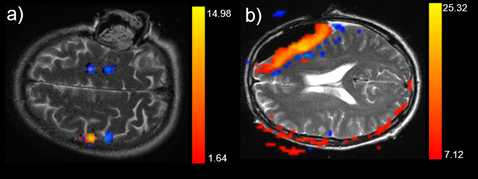
Figure 1: Representative examples of the activation of
the motor cortex (a) and false activation patterns in the resection cavity (b)
overlaid on T2 images.
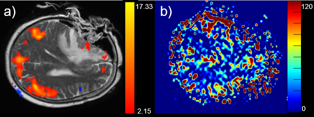
Figure 2: Example of the default mode network (a)
overlaid on a T2 image and the corresponding ASL perfusion (b). Note that in
the frontal area (medial prefrontal cortex and anterior cingulate cortex) no
default mode activation is visible as compared to the standard situation. The
inferior parietal cortex and cingulate cortex as well as the precuneus are visualized
as expected.