3380
Optimization of simultaneous multislice (SMS) technique for submillimeter whole brain functional MRI at 7T1MR, GE Healthcare, Waukesha, WI, United States, 2GE Healthcare, Waukesha, WI, United States, 3GE Healthcare, Buc, France
Synopsis
Through the optimization of simultaneous multislice (SMS) technique, a 0.8mm3 whole brain resting state fMRI data with minimum distortion was acquired to show the benefit of doing fMRI study at ultra-high field.
Introduction
The benefit of doing functional MRI at ultra-high field (>= 7T) are the increased signal-to-noise ratio (SNR) and higher BOLD sensitivity. While the temporal resolution is crucial to capture the BOLD dynamics in fMRI experimental design, spatial resolution is beneficial for better localization of activation areas and differentiation of cortical layers, for example and there is always trade-off to be made between spatial and temporal resolution. The simultaneous multislice (SMS) technique which shortens the repetition time (TR) and consequently the scan time by roughly a factor equal to the number of simultaneously excited slices, without significant signal-to-noise ratio (SNR) penalty, has been widely adopted for improving temporal resolution in fMRI especially in the domain of neuroscience research (1, 2). Blipped-Controlled Aliasing in Parallel Imaging (blipped-CAIPI) (3) is used in SMS acquisition to reduce the noise amplification, in-plane signal aliasing.With the improved SNR at 7T, higher spatial resolution such as <=1mm3 isotropic voxels fMRI can be obtained in reasonable imaging times while maintaining high temporal resolution with the help of simultaneous multislice (SMS) EPI technique. However, there are some technical challenges for echo-planar imaging at ultra-high field, such as increased susceptibility artifacts near the frontal sinuses and skull base, producing spatial distortion and signal drop out. It is desirable to combine SMS with high in-plane parallel imaging acceleration to reduce the echo-train length and distortion. But this can lead to a high SNR penalty. The slice shift in blipped-CAIPI SMS technique is usually set to a fixed fraction of field of View (FOV), experimentally predefined for a coil and hyperband factors.
Here, an offline optimization of slice FOV shift factor based on experimental setup such as coil sensitivity map, FOV, number of slices, SMS factor and in-plane acceleration factor is used to acquire a fMRI whole brain resting state data at 0.8mm3 isotropic resolution with a TR of 2.84 second.
Method
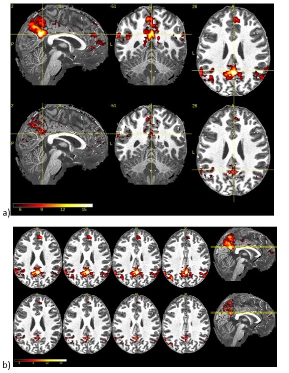
One human dataset was acquired on GE Signal 7T (GE Healthcare, Waukesha, WI, USA) under the guidelines of Institutional Review Board using a NOVA single-channel transmit, 32-channel phased-array receive head coil (Nova Medical Inc., MA, USA). The T1-weighted anatomical image was acquired using 3D MPRAGE sequence, scan parameters are TR of 3.409s, TE of 3.9ms, TI of 1.1s and FOV of 21.6cm with 0.7mm3 isotropic pixel size. For the resting state fMRI scan, the product SMS EPI pulse sequence was used with following parameters: SMS factor of 5, in-plane acceleration of 3, TR of 2.84s , TE of 30ms, flip angle of 80 degree, FOV of 25.6cm, slice thickness of 0.8mm, and in-plane resolution of 0.8mmx0.8mm. Total 250 volumes were acquired; total scan time is about 12minutes. The CAIPI shift was select as ½ of FOV through optimization based on g-factor estimation according to FOV, resolution, coil sensitivity map, SMS factor, in-plane acceleration factor (4). The larger FOV of 25.6cm was selected to further reduce the estimated g-factor for SNR improvement using the same offline optimization.Resting state fMRI data was processed by SPM12 software using this pipeline: 1) simple realignment, slice timing correction; 2) co-registration of EPI to 3D-T1w, co-registration of 3D-T1w to T1 MNI template, normalization of 3D-T1w, apply transformation to EPI; 3)application of brain mask, detrending and bandpass filtering [0.008 0.1] Hz, residualisation using anatomical CompCor (5), motion parameters and their time derivatives; 4) smoothing (FWHM = 2mm and 4mm). A seed-based analysis using a 10mm diameter sphere in the PCC (centered at MNI Coordinate [2 -54 26]) to extract DMN (default mode network). Seed time course are extracted before smoothing and used at regressors of interests in GLM with the smoothed data.
Results
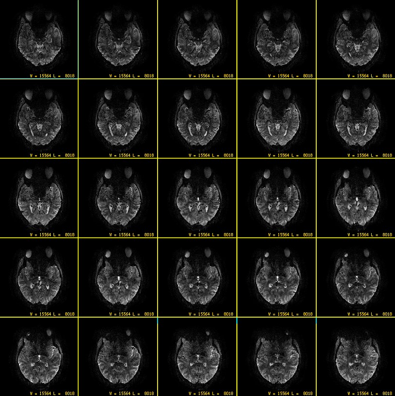
Figure 1 is the axial display of multi slice resting slice EPI images, there are minimum EPI distortions due to the higher in-plane acceleration. By increasing FOV from typical 21-22cm to 25.6, the average whole brain tSNR increased 52% as predicted by offline g-factor estimation.PCC Seed-based default mode network Result with 4mm (top) and 2mm (bottom) smoothing are shown in Figure 2. The DMN map with 2mm smoothing demonstrated that submillimeter whole brain fMRI data provided better localization of activation areas and differentiation of cortical layers.
Conclusion
The proposal optimization of SMS technique reduced the image artifact and improved tSNR for fMRI scan at 7T.Acknowledgements
No acknowledgement found.References
1. Simultaneous Multislice (SMS) Imaging Techniques, Markus Barth, Felix Breuer, Peter J. Koopmans, David G. Norris, and Benedikt A. Poser, Magn Reson Med. 2016; 75:63–81 .
2. Multiband multislice GE-EPI at 7 tesla, with 16-fold acceleration using partial parallel imaging with application to high spatial and temporal whole-brain fMRI, Moeller S, Yacoub E, Olman CA, Auerbach E, Strupp J, Harel N, Uğurbil K, Magn Reson Med. 2010; 63(5):1144-1153
3. Blipped-Controlled Aliasing in Parallel Imaging (blipped-CAIPI) for simultaneous multi-slice EPI with reduced g-factor penalty, Kawin Setsompop, Borjan A. Gagoski, Jonathan R. Polimeni, Thomas Witzel, Van J. Wedeen, and Lawrence L. Wald, Magn Reson Med. 2012 May ; 67(5): 1210–1224.
4. SENSE: sensitivity encoding for fast MRI, K P Pruessmann, M Weiger, M B Scheidegger, P Boesiger, Magn Reson Med. 1999 Nov;42(5):952-962.
5. A Component Based Noise Correction Method (CompCor) for BOLD and Perfusion Based fMR: Yashar Behzadi, Khaled Restom, Joy Liau, and Thomas T. Liu, Neuroimage. 2007;37(1): 90–101.
Figures