3359
Commercially available astriction cotton as a anisotropic DTI phantom: Comparisons with a hand-bundled fibers and a glass capillary plate1Kyoto Prefectural University of Medicine, Kyoto, Japan, 2National Institute of Radiological Sciences, Chiba, Japan, 3Kyoto Prefectural University of Medicine Hospital, Kyoto, Japan
Synopsis
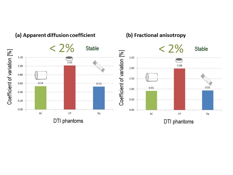
We compared DTI measures among three different anisotropic diffusion phantoms: a commercially available astriction cotton; a glass capillary plate; a hand-bundled polyethylene fibers. The purpose of this study was to examine whether the cotton is useful as an anisotropic diffusion phantom. Differences in ADC and FA were evaluated by comparing coefficients of variation (CVs). No significant difference was seen for the largest eigen value: 2.06 - 2.14 x 10⁻³ mm2/sec. Clear differences in FA were seen among the three DTI phantoms (p = 0.0079). CVs from five acquisitions for ADC and FA from every phantoms were all less than 2%.
INTRODUCTION
In general, a large data set gives larger statistical power for study results. While collecting large data sets is appropriate for research, accomplishing this using only a single magnetic resonance (MR) scanner is not possible. As a result, multi-scanner studies are often needed to fulfill the statistical requirements of MR studies. Nevertheless, intrinsic differences among scanners have been recognized as inevitable. The Quantitative Imaging Biomarkers Alliance (QIBA) was established to address these intrinsic differences and obtain statistically meaningful results from multi-scanner studies 1. Isotropic diffusion has already been discussed by the QIBA 2. However, anisotropic diffusion has not been included among QIBA endeavors because of the absence of appropriate anisotropic diffusion phantoms. A suitable phantom needs to satisfy these points: stable measurement of values from diffusion tensor imaging (DTI); cost-effectiveness; and availability. Anisotropic diffusion phantoms have been used, including the solid capillary type 3-5 and bundled fiber type 5, 6. However, these phantoms have various drawbacks. The solid capillary type shows only hindered diffusion and is expensive, and unsuitable for widespread use. On the other hands, most bundled fiber-type phantoms are handmade, and preparation of multiple identical pieces for industrial application is difficult. This study compared the DTI values of apparent diffusion coefficient (ADC), eigen value (λ), and fractional anisotropy (FA) among three different anisotropic diffusion phantoms: commercially available astriction cotton (AC)7; a glass plate (CP)5; and hand-bundled polyethylene fibers (Dy)5. The purpose of this study was to examine whether AC is useful as an anisotropic diffusion phantom for multi-scanner studies.MATERIALS & METHODS
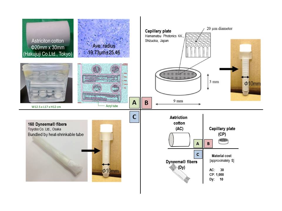
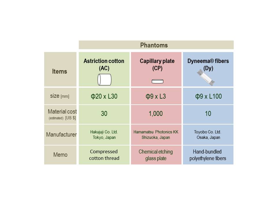
Phantoms: AC (30 mm in length, 20 mm in diameter; Hakujuji Co., Tokyo, Japan) in acryl tubes (diameter, 25 mm) was arranged in layers of parallel tubes arranged perpendicular to the underlying layer (Figure 1a). The phantom was 12.5 cm wide x 17.0 cm long x 12.0 cm high (polyethylene container) 7. The capillary glass plate (Hamamatsu Photonics K.K., Shizuoka, Japan) was 9 mm in diameter and 3 mm thick, with uniformly spaced micro-pores (diameter, 20 μm). Twenty plates were stacked in a plastic tube with ultra-pure water (Figure 1b) 5. The polyethylene fibers (Dynnema®; Toyobo Co., Osaka, Japan) were bundled together with a thermal shrinkage tube 5. These bundled fibers were placed in a plastic tube with ultra-pure water (Figure 1c). Table 1 summarizes the features of the phantoms.MR scanner: This study used a 3-T MRI system (MAGNETOM Verio 3T; Siemens Healthcare K.K., Erlangen, Germany) equipped with a 12-channel head coil. The phantoms were placed in parallel with the Z-axis direction of the MRI gantry.
Data acquisition: DTI was performed using b values of 0 and 1000 s/mm2 in 10 different diffusion-encoding directions. The field of view was 240 x 240 mm. DTI was acquired using an EPI technique with a matrix of 120 x 108. Of note, diffusion time was automatically selected by the MRI system for this additional scan, but was not displayed on the user interface as is usual for clinical machines. Other parameters were: TR = 6300 ms; TE = 92 ms; average = 3; number of b0 images = 1; number of readout segments = 5; resolution = 2.0 × 2.0 × 2.0 mm; room temperature, approximately 22°C; and total scan time = 4 min. Five acquisitions were performed for each phantom.
DTI analysis: DTI analysis was performed with Diffusion Toolkit/TrackVis version 0.5.2.28, and hand-drawn regions of interest (ROIs) based average diffusion metrics were calculated by 3D slicer 9. Evaluation of stability: The stability of diffusion metrics (ADC and FA) of the phantoms was evaluated by comparing coefficient of variation (CV) as CV [%] = standard deviation / mean x 100. When CV was < 5%, the measurement was considered homogeneous 10.
Statistics: Comparisons between DTI measurements from different phantoms were performed using the Wilcoxon rank-sum test (Matlab®; The MathWorks, Natick, MA). The difference was evaluated as statistically significant for Values of p < 0.05 were evaluated as statistically significant.
RESULTS
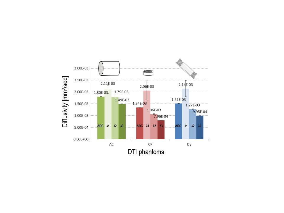
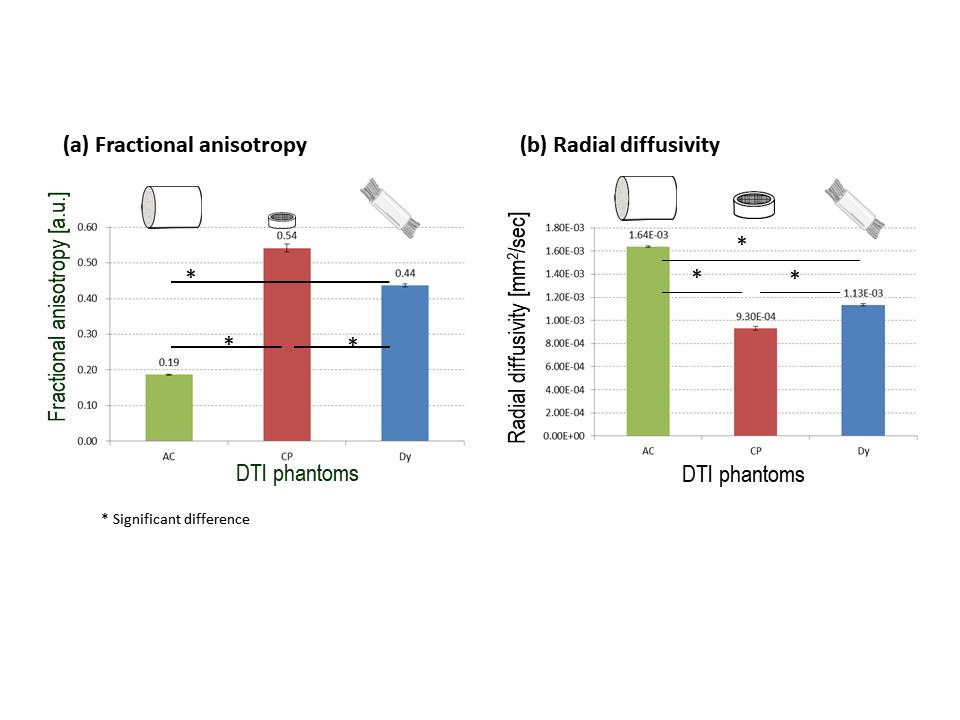
Diffusivity: In Figure 2, the average ADC of three different phantoms were 1.34 – 1.80 x 10⁻³ mm2/sec (p < 0.05). In contrast, no differences in largest eigen value were seen among the three DTI phantoms: 2.06–2.14 x 10⁻³ mm2/s (p: AC vs. CP = 0.15, CP vs. Dy = 0.095, Dy vs. AC = 0.15).Anisotropic diffusion: FA values of the three phantoms were 0.19–0.54 (Figure 3a). Clear differences in FA were seen among the three DTI phantoms (p = 0.008), attributed to significant differences in radial diffusivity (p = 0.008; Figure 3b).
Inter-5-scan difference: CVs of ADC (Figure 4a) and FA (Figure 4b) for the phantoms were all <2%. Scan-rescan diffusivity and anisotropy were thus stable for all three phantoms.
DISCUSSION & CONCLUSION
CP and Dy showed high FA and are considered appropriate as anisotropic diffusion phantoms. However, chemically etched glass plates are expensive and unsuitable for widespread use. Since Dy is handmade, preparation of multiple identical pieces for industrial application is difficult. In contrast, AC is a mass-produced industrial material and inexpensive. AC is expected to solve the drawbacks of CP and Dy. AC shows relatively low FA, but the range of values observed in the living brain 11.Acknowledgements
This work was supported by JSPS KAKENHI Grant Number JP17K10413.References
1. QIBA, https://www.rsna.org/qiba/.
2. http://qibawiki.rsna.org/images/1/1b/QIBA_IMI_August_7_2013-M_Boss.pdf.
3.Yanasak N et al., Magnetic Resonance Imaging. 2006; 24: 1349–1361.
4.Oida T et al., Magn Reson Med Sci. 2011: 10(2); 121-128.
5. Tachibana et al., Magn Reson Med Sci. 17(2018), pp.251-258.
6.Lemberskiy G et al., NMR in Biomedicine. 2017;30:e3708
7. Sakai et al., Proc. Intl. Soc. Mag. Reson. Med. 24 (2016), 2032.
8. Diffusion toolkit, https://www.nitrc.org/projects/trackvis/.
9. 3D slicer, https://www.slicer.org/
10. Kamagata et al., Magn Reson Med Sci. 2015; 14: 227-233.
11. Alexander AL et al., Neurotherapeutics, 2007; 4: 316-329.
Figures