3355
1:1 Scale Agar-Agar Paramagnetic Phantom for Brain and Cervical Spine MRI
Elif Aygun1,2, Ahmet Rahmetullah Cagil1,2, and Emine Ulku Saritas1,2,3
1Department of Electrical and Electronics Engineering, Bilkent University, Ankara, Turkey, 2National Magnetic Resonance Research Center (UMRAM), Bilkent University, Ankara, Turkey, 3Neuroscience Graduate Program, Bilkent University, Ankara, Turkey
1Department of Electrical and Electronics Engineering, Bilkent University, Ankara, Turkey, 2National Magnetic Resonance Research Center (UMRAM), Bilkent University, Ankara, Turkey, 3Neuroscience Graduate Program, Bilkent University, Ankara, Turkey
Synopsis
Phantoms doped with paramagnetic materials are commonly used for quality assessment of MRI protocols and image reconstruction techniques. These phantoms are typically prepared using cylindrical containers and mimic the T1 /T2 characteristics of human tissue. In this work, we propose a 1:1 scale human head and neck agar-agar phantom prepared using a 3D human model. The phantom mimics human tissue characteristics while incorporating the skull and cervical vertebrae. The MRI images show that the phantom closely matches human anatomy, and the susceptibility artifacts in EPI images successfully mimic those seen during in vivo imaging of the brain and spine.
Introduction
Susceptibility artifacts are common in echo planar imaging (EPI) sequences. In vivo imaging of the spinal cord particularly suffers from these artifacts due to susceptibility differences between the various tissue types found in the neck.1 Standard paramagnetic phantoms that are built for quality assessment in MRI are prepared in cylindrical containers. In this work, we propose a 1:1 scaled agar-agar paramagnetic gel phantom that reflects human head and neck anatomy for quality assessment of MRI protocols.Methods
3D DesignThe 3D design was performed in Autodesk FUSION 360 and Autodesk Meshmixer. Two reference models were used for the body2 and the skull and spine3. These models were altered to include skull, cervical spine, thoracic spine, and first two lumbar vertebrae. The body was scaled to match the proportions of an average human of 170 cm height, and the design was cut horizontally under the chest level to have a final height of 44.2 cm, with an 8-mm wall thickness. For the insertion of skull and spine, the design was separated into two interlocking parts 12.2 cm below the top of the head, with 4-mm thick indents created reciprocally on each side to ensure a tight fit (see Fig. 1). The teeth were removed from the model to reduce complexity, and the spine was altered to have a flat surface under the second lumbar vertebrae. Lastly, the skull-spine system was re-scaled to fit into the body (see Fig. 2 and Fig. 3). The body was 3D printed using PETG with 96% infill to ensure water tightness, and the skull-spine system was 3D printed using PLA. The parts were attached to each other using super glue and the exterior interlocking parts were melted together to seal the gaps. Finally, XTC-3D epoxy was used to make the model watertight. Shown in Fig. 2(a), the final size of the phantom was 30.5 x 24.7 x 44.2 cm, with a volume of 14L.
Agar-Agar Gel Preparation
The paramagnetic agar-agar gel was prepared to mimic human white matter T2, reported as 69 ms at 3T. 4,5 CuSO4 was chosen as the paramagnetic agent and NaCl was added to increase conductivity to be similar to that of human tissue.6 Agar-agar gel mimics soft human tissues when prepared at appropriate concentrations, where the solidification performance of the gel also depends on concentration. To determine the appropriate concentration, 3 different samples were prepared with 1%,1.5%, and 2% agar-agar in 500 mL containers. The samples were imaged with T2 -weighted turbo spin echo (TSE) sequence with a series of TE values. The resulting images shown in Fig. 4(a) were used to compute T2 via fitting to an exponential signal decay curve.4 Based on these results, the agar-agar concentration was chosen as 1.5% to achieve slow solidification without saturation. Based on the measured T2 values from Fig. 4, the required CuSO4 concentration to match white matter T2 was adjusted as 4.25mM. For this combination of agar-agar and CuSO4 concentrations, the T2 was measured as 61.9 ms, closely matching the target 69 ms.
The gel was prepared in 1L batches with 1.5% agar-agar powder, 4.25 mM of CuSO4, and 0.2% NaCl.7 For each batch, 15 grams of agar-agar and 2 grams of NaCl were mixed with 1L of distilled water, stirred until boiling, and simmered for 2 minutes.7 Then, 692 mg of CuSO4 was added while stirring slowly to avoid formation of air bubbles that disrupt the homogeneity of the gel. Mixed every 3 minutes, the gel was allowed to cool down to 55°C to avoid deformities in the phantom, considering the 60°C heat tolerance of PETG. This entire process was repeated in 1L batches until the phantom was filled, with the phantom shaken repetitively to avoid air bubbles. To allow air contact for complete solidification, the phantom was kept uncovered at room temperature for 24 hours before sealing closed.
MRI Acquisition
The phantom was imaged on a Siemens Magnetom 3T scanner with an 8-channel spine array coil. EPI images were acquired in the axial and sagittal planes with 330 mm and 400 mm FOV, respectively. Common parameters were TE= 28 ms, TR=6500 ms, voxel size =2.5x2.5x5 3 ,16 averages, GRAPPA factor of 4. TSE images were acquired in the axial and sagittal planes with 300 mm and 400 mm FOV, respectively, with TE= 12 ms, TR= 3500 ms, slice thickness = 3 mm.
Results
The final phantom is shown in Fig. 3(b), with the MRI images in Fig. 5. TSE image in the sagittal and axial plane show that the phantom closely matches the human anatomy, while the EPI images show susceptibility artifacts prominent in regions such as the neck where the susceptibility is known to vary rapidly.Conclusion
We developed a 1:1 scaled agar-agar gel MRI phantom that reflects human anatomy while matching tissue characteristics. The MRI images show that the phantom closely matches human anatomy, and the susceptibility artifacts in EPI images successfully mimic those seen during in vivo imaging of the brain and spine. The phantom will be utilized in the quality assessment of the MRI protocols and image reconstruction techniques in the brain and the spine.Acknowledgements
This work was supported by the Scientific and Technological Research Council of Turkey (TUBITAK 117E116).References
- Cohen-Adad J and Wheeler-Kingshott C. Quantitative MRI of the spinal cord. San Diego, Calif.: Academic Press. 2014; 91-103.
-
Abodanyal. Fdx54mtvuz28- Final Base Mesh. Sketchfab. https://sketchfab.com/3d-models/fdx54mtvuz28-final-base-mesh-ad130e51f77c4bf2aa730684c67fc69e. Accessed August 13, 2020.
-
Hamer S. Human Anatomy V4 - Male (Life
Sciences Japan, Body-Parts3D Source). 3D CAD Model Collection | GrabCAD
Community Library. https://grabcad.com/library/human-anatomy-v4-male-life-sciences-japan-bodyparts3d-source-1. Accessed October 12, 2018.
- Thangavel K. and Saritas EU. Aqueous paramagnetic solutions for MRI phantoms at 3 T: a detailed study on relaxivities. Turkish Journal of Electrical Engineering & Computer Sciences.2017;25:2108-2121
- Stanisz GJ, Odrobina EE, Pun J et al. T1, T2 relaxation and magnetization transfer in tissue at 3T. Magn Reson Med. 2005;54(3):5017-12.
- Yoshida A et al. Development of a phantom compatible for MRI and hyperthermia using carrageenan gel—relationship between T1 and T2 values and NaCl concentration. International Journal of Hyperthermia. 2004;20(8):803-814.
- Hamhaber U, Grieshaber, Nagel J, et al. Comparison of quantitative shear wave MR-elastography with
mechanical compression tests. Magnetic Resonance in Medicine. 2002;49(1):71-77.
Figures
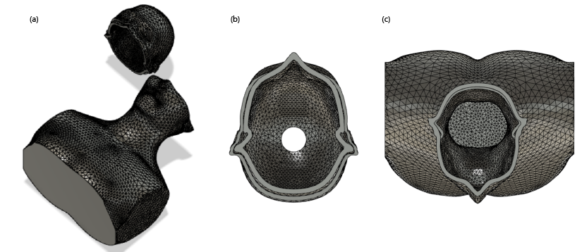
Figure 1. (a) The 3D model of
the phantom was designed in Autodesk Fusion 360 based on a human model. To place
the spine and the skull inside of the phantom, the model was divided into two
parts: (b) a section of the top of the head with 12.2-cm height and (c) the
rest of the model. Indents of 4-mm thickness and 2-cm length were created on
each side to ensure a tight-fitting interlock of the two parts.
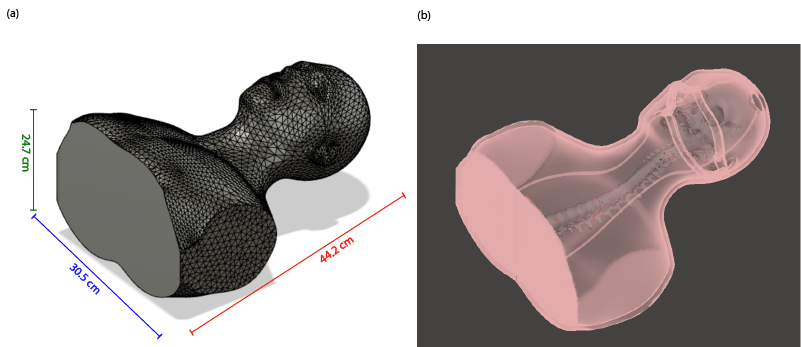
Figure 2. (a) The phantom
dimensions were 30.5 x 24.7 x 44.2 cm3 with 8-mm wall thickness. (b)
The spine and the skull were placed inside of the phantom, aligned post-print with
the center of the phantom. The spine was attached to the skull from the Atlas
bone with plastic cable ties and super glue, and was fixed to the bottom of the
phantom using hot glue. These fixations ensured that the spine remains in its
intended place during the filling of the agar-agar gel.
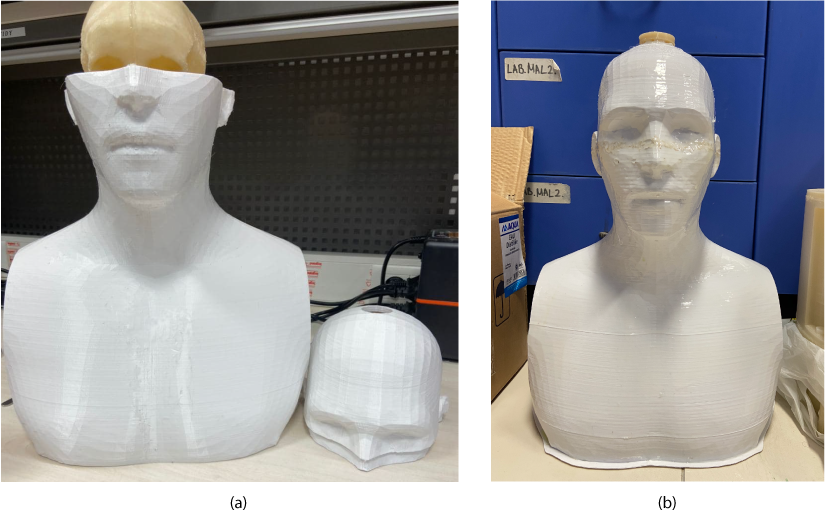
Figure 3. (a) The skull and
spine system was placed inside the phantom post-3D-print. (b) Then, the top part of the head was superglued to the bottom part, and the phantom was sealed by melting the PETG
material in the seams of the interlock. The model was coated with XTC-3D epoxy
to ensure a watertight design. The phantom was then filled with agar-agar gel
and left uncovered at room temperature for 24 hours. After the gel solidified,
the opening on the top was sealed to preserve the phantom.
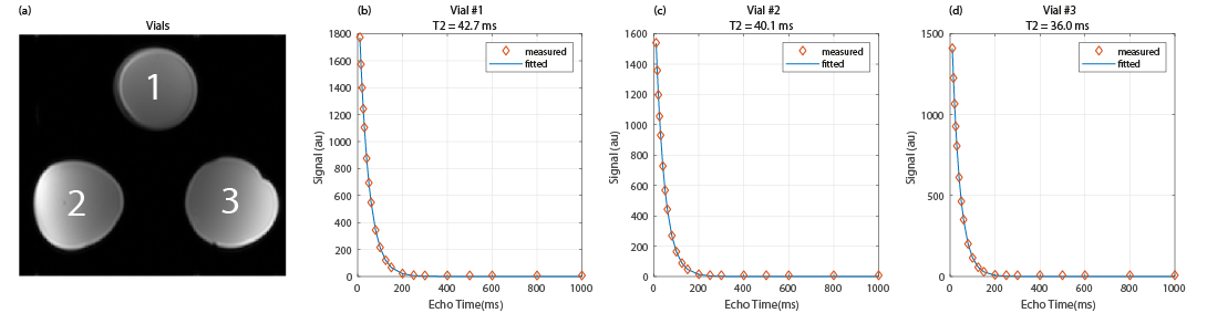
Figure 4. Three samples with 1%,
1.5%, and 2% agar-agar concentrations were prepared with equal amount of CuSO4
to determine the effect of agar-agar on T2 . (a) The samples were
imaged with a TSE sequence with a series of TE values. (b)-(d) T2 for each sample was computed via fitting to an
exponential signal decay curve. As a result, 1.5% agar-agar concentration was
chosen to prepare the phantom, and CuSO4 concentration was adjusted
as 4.25 mM to reach a final measured T2 value of 61.9 ms.
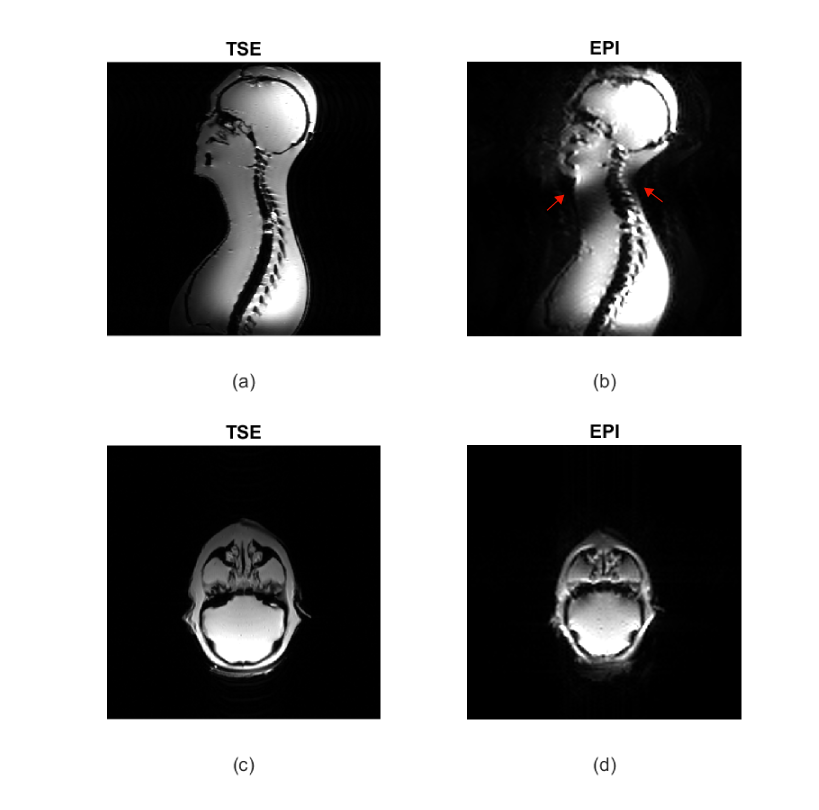
Figure 5. MRI images acquired
with TSE and EPI sequences. (a-b) Sagittal images of the whole phantom and the
spine. The susceptibility artifacts are seen on the back and front of the neck for
EPI (red arrows) successfully mimick the problems seen during in vivo spine
imaging. (c-d) The brain in the axial plane also shows susceptibility artifacts,
similar to the ones seen during in vivo brain imaging.