3325
Single Half-Cylinder B0 Shim Coil for the Knee1Case Western Reserve University, Cleveland, OH, United States, 2Quality Electrodynamics, Mayfield, OH, United States
Synopsis
The void under the knee of a supine patient leads to localized B0 magnetic field distortions due to air-tissue susceptibility differences. To cancel this local artifact, low-order harmonic shims are inadequate, but a single local shim coil can produce higher harmonic suppression. A half-cylinder version wrapped on the outside of the RF coil has been constructed and used to obtain in vivo data consistent with the magnetic field simulations. The detailed results support the possibility of an effective knee shimming approach.
Introduction
Improvements in SNR carry with it an increased need to reduce the effects of susceptibility artifacts, especially at higher field strengths. Large discontinuities occur for the air-tissue interface, the basis for a concern in knee imaging, particularly for an elderly patient population. An issue for the aged and the injured may be an inability to straighten fully the leg during imaging, which exacerbates the artifact under the knee. We can address the specific air-body discontinuity, which gives rise to a localized spatial change in the B0 field, with local shim coils. Such coils produce correspondingly high-order spherical harmonics, unlike those available from the built-in low-order spherical harmonic shims of the main magnet. Local shims have therefore been developed with multiple coils as well as single coil patterns, generally relying on field-driven maps to characterize the artifacts, and look to be effective for a variety of applications from brain to large organs such as the liver.1-6In view of smaller attention being paid to the knee, we consider a single shim “half-coil” addressed to the above-described problem for knee imaging of the general patient population. We construct and test a new half-cylinder coil wrapped around an RF knee coil. The coupling of this coil to the surrounding system conductors is reduced for such a small footprint. Our experimental results are consistent with simulations based on the design coil current pattern.Methods
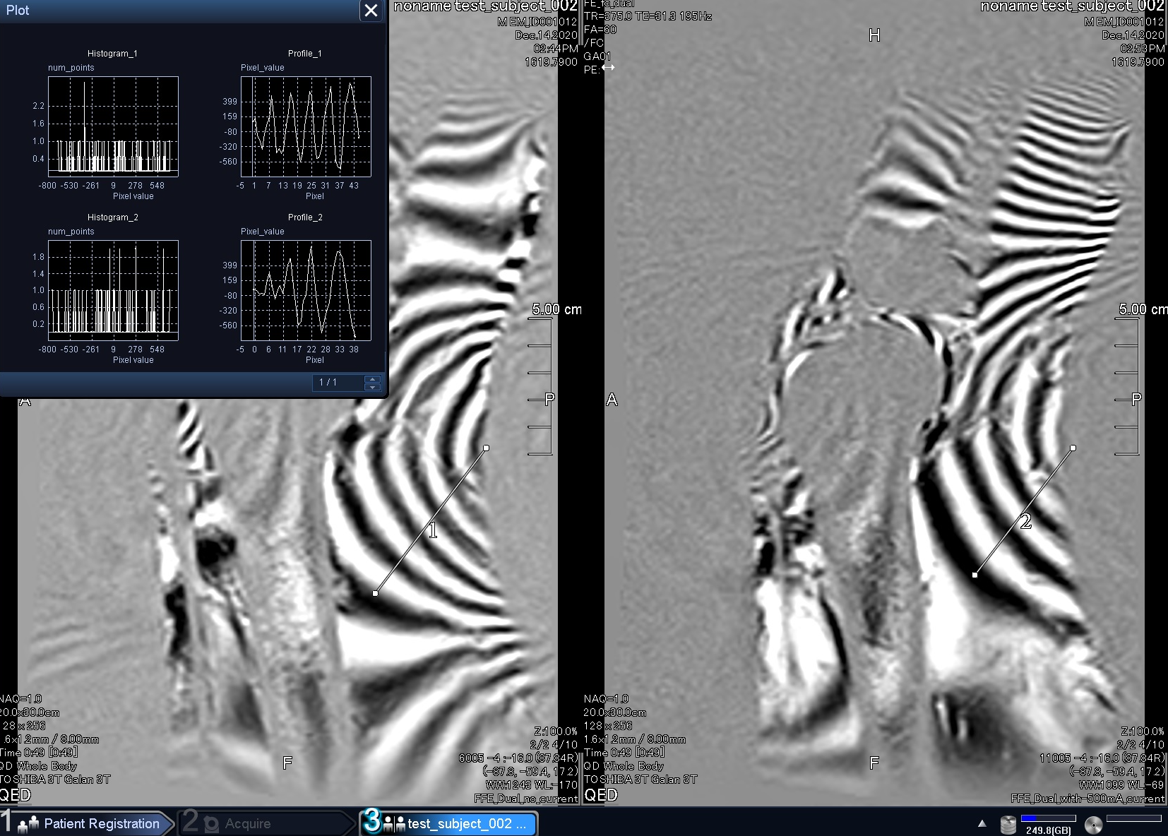
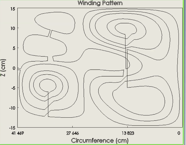
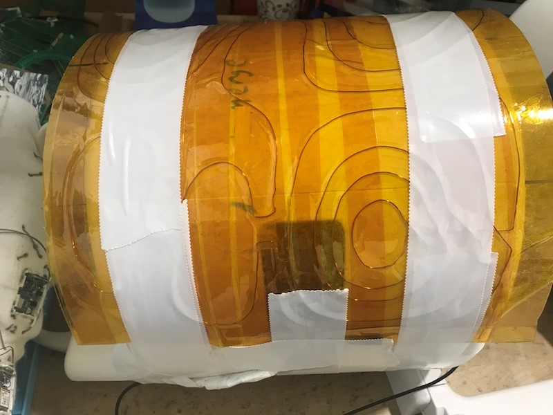
Phase maps are found with dual-echo RF pulse sequences (Sequence: FE2D, FC_Dual, TE1:15ms,TE2:31.3ms,, TR:375ms, FA: 60degrees, FOV: 20x30cm) Fig. 1 shows the bent-knee phase images of a healthy volunteer (6’5” and 180lbs) exposing the targeted artifact on the interior surface. The images were obtained with a Canon Galan 3T whole-body system with a 16ch Tx/Rx knee coil. To find the target field for design purposes, a 3D B0 map was divided into ten y-z sagittal slices along the x-axis. In a generalized Turner approach7 applied to partial cylinders8, the cost functional is $$$L= E + λW$$$ , in which E is the inhomogeneity quadratic remaining after shimming and W is an integral of the square of the current density over the coil surface. W controls the power consumption and the regularization parameter λ permits a trade-off between the remaining error and the wire current. The L extremum yields a linear solution for the surface current density in terms of sinusoidal basis functions of a cylindrical geometry. We aim for a proof-of-principle demonstration in the present paper by wrapping the shim around the outside of the RF knee coil (length 30cm, radius 13.2cm). The density is discretized via a stream function into closed-loop islands. To obtain a single-wire coil, the closed-loops are connected step-wise in spiral fashion.Results
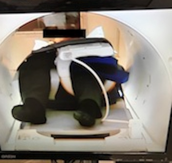
By dialing λ, a sweet-spot is found between the number of loops and the current, and an optimized current pattern with spiraled connections is shown in Fig.2. It corresponds to a current of 500mA, with a relatively uncrowded set of concentric loops and islands on the half cylinder surface. The shim half-coil was constructed and is pictured in Fig.3; Fig. 4 is a photograph of the volunteer inside the scanner bore. Fig.1 displays the phase map in the center sagittal plane (x=0) before the shimming (left-hand side) and after shimming (right-hand side). Now the B0 map used for this optimization corresponds to a different constraint, but the result can still be used for a proof-of-principle and for quantitative testing. By counting the black-white transitions along the indicated reference lines (0.25ppm per π transition), for example, we see that they have dropped approximately from five to four (20%). This small reduction is expected for the coil because its target was not at the knee-bending center but 5cm above it. We thus have two confirming features from Fig. 1. The first is the 20% reduction is consistent within errors (10%) at the center and the region above the center reveals an increase in transition number (phase stripes). This coil in fact generates an artifact where there was none. Note that it can be seen by eye the local coil effect drops off sufficiently away from the ROI.Discussion
The results for a single shim half-cylinder coil wrapped around the RF coil agree with simulations based on the given current pattern. This is evidence for the usage of half-coil functional methods to significantly reduce the local field inhomogeneity in a representative volunteer, even with a bent-knee posture. While the half-coil used was designed for a whole-body transmit experiment and a different B0 map, its effect on the artifact and phase map in the present experiment could be compared to the coil simulation. The reduction in artifacts in the center and the production of artifacts above center were in agreement with theory.Conclusion
A knee shim approach can take advantage of the fact that the specific susceptibility area of interest behind the knee is relatively generic over a large fraction of the patient population. Based on the experimental results found, a half-coil designed for the appropriate scanning setup and hence the appropriate B0 map can be an effective knee artifact management for the future.Acknowledgements
Support for this work has been provided by the Ohio Third Frontier Program.References
1. L. van den Wildenberg et al. B0 shimming of the liver using a local shim coil array at ultra-high-field MRI. ISMRM 2020. Abstract 4220.
2. S. E. Ungersma, H. Xu, B. A. Chronik, G. C. Scott, A. Macovski, and S. M. Conolly. Shim design using a linear programming algorithm. Magnetic Resonance in Medicine, 52, 619-627, 2004.
3. J. P. Stockmann and L. L. Wald. In vivo B0 field shimming methods for MRI at 7T. Neuroimage, 168, 71-87, 2018.
4. B. P. Meneses and A. Amadon. A fieldmap-driven few-channel shim coil design for MRI of the human brain Physics in Medicine and Biology, accepted, PMB-110883.R1, 2020.
5. C. Liao et al. Distortion-free, submillimeter-isotropic-resolution diffusion MRI with gSlider BUDA-EPI and multi-coil dynamic B0 shimming. ISMRM 2020. Abstract 0978.
6. C. Juchem et al. Dynamic multicoil technique (DYNAMITE) MRI on human brain. Magn Reson Med, 84, 2953–2963, 2020.
7. Turner, Robert. Minimum inductance coils. Journal of Physics E: Scientific Instruments 21.10 (1988): 948.
8. Fan, Mingdong. Three Initiatives Addressing MRI Problems. Case Western Reserve University, Ph.D. thesis March, 2020.
Figures