3254
Substantia Nigra Abnormalities in Early Parkinson’s Disease Patients using Convolutional Neural Networks in Neuromelanin MRI1CENIR, ICM Paris, Paris, France, 2Paris Brain Institute (ICM), Sorbonne University, UPMC Univ Paris 06, Inserm U1127, CNRS UMR 7225, Paris, France, 3ICM Team “Movement Investigations and Therapeutics” (MOV’IT), Paris, France, 4Department of Neuroradiology, Pitié-Salpêtrière Hospital, AP-HP, Paris, France, 5INSERM, Clinical Investigation Center for Neurosciences, Pitié-Salpêtrière Hospital, Paris, France, 6Biogen Inc., Cambridge, MA, United States, 7Department of Neurology, APHP, Pitié-Salpêtrière Hospital, Paris, France
Synopsis
There is a need of accurate imaging biomarkers of dopaminergic cell neurodegeneration to facilitate drug trials in Parkinson’s disease (PD). PD demonstrates neurodegenerative substantia nigra pars compacta (SNc) changes that can be detected efficiently using neuromelanin-sensitive MRI. Characterizing neuromelanin signal variations using manual SNc segmentation is an operator-dependent and time-consuming task. Hence, in this cross-sectional, observational, case-control study, we investigated neuromelanin SNc abnormalities in the early PD patients using convolutional neural network-based fully automatic segmentation of SNc. We found a highly significant difference in SNc volume and signal intensity between early PD and healthy volunteers.
Introduction
Parkinson disease (PD) impacts 2–3% of the population ≥65 years of age and is characterized by the progressive loss of dopaminergic neurons in the substantia nigra pars compacta (SNc) resulting in striatal dopamine depletion1-3. Motor symptoms in PD start when the dopaminergic neuronal loss reach around 30% to 60%4,5. SNc dopaminergic neurons contain a neuromelanin pigment6 that is visible using neuromelanin-sensitive imaging6-8.Studies have reported reduced size and signal intensity of the SNc in PD patients using neuromelanin-sensitive imaging8-11 and were able to differentiate the PD patients from the healthy volunteers (HV) using semi-automated and manual segmentation of SNc for computing the volume and signal of SNc11-12. However, such methods are time-consuming and prone to substantial inter-individual variability across raters. Automated segmentation approaches can be less prone to errors and may lead to a higher consistency in SNc regions of interest (ROI) assessment13-15.
Deep learning is a machine learning class of artificial intelligence (AI), which includes neural networks with various layers that is widely used in image recognition and segmentation tasks16. Convolutional neural networks (ConvNet) use a simple artificial neural network architecture that has proven to perform far better than common AI tasks16,17,18.
In this study, we investigated the neuromelanin signal changes in PD patients using ConvNet-based fully automatic segmentation of SNc, compared the measurements to the manual SNc segmentations and explored its potential value as a biomarker of disease modification in clinical neuroprotective trials.
MATERIALS AND METHODS
PD patients and healthy volunteers were prospectively included in the ICEBERG study conducted at the Paris Brain Institute. Subjects were scanned using a 3 T PRISMA scanner (Siemens) and a 64-channel receive head only coil. 3D T1-w images were acquired using a sagittal Magnetization Prepared 2 RApid Gradient Echo (MP2RAGE) with a 1-mm isovoxel size19 and neuromelanin-sensitive images were acquired with the following parameters: with TR/TE/flip angle: 890ms/13ms/180°, 3 averages, voxel size: 0.4×0.4×3 mm3, acquisition time (TA): 6:55 min.Manual segmentations of the SNc ROI were performed on the neuromelanin-sensitive images. The SNc was defined as the hyperintense area dorsal to the cerebral peduncle and ventral to the red nucleus (Figure 1). A background region was also manually traced including the tegmentum and superior cerebral peduncles . These manual segmentations were performed by two independent expert examiners blinded to the status of the subject using the FreeSurfer viewer similar to previous study20.
Deep learning segmentations were performed using U-net architecture of ConvNet21 by employing ‘adam’ method (Figure 1). From the pool of 140 manually segmented images of PD patients and HV, a random training dataset of 42 images and 6 validation images were prepared for the neural network.
The SNc volumes, corrected volume (Cvol) by dividing SNc volumes by total intracranial volume to correct for individual head sizes, signal-to-noise ratio (SNR) and contrast-to-noise ratio (CNR) by normalizing the mean signal in SNc relative to the background signal were calculated20. Furthermore, we performed a two-way multivariate GLM–ANOVA with Status (PD, HV) as between-group factor while adjusting for age and sex. Inter and intra-observer variability was estimated using Dice-coefficient. Pearson’s correlation coefficients were calculated between SNc measurements and clinical scores. To adjust for multiple comparisons, an approximate multivariate permutation test was conducted22. The effect of levodopa equivalent daily dose (LEDD) in PD patients were also calculated (Table 1).
RESULTS AND DISCUSSION
Ninety-nine early PD patients (mean disease duration = 1.5 ± 1.0 years) and 41 HV were analyzed, with no significant difference in age between the groups but having a larger proportion of males among patients (χ2=7.630, p=0.005) (Table 1).For both ConvNet and manual methods, we found a highly significant difference in all SNc measurements of Volume, Cvol, SNR, CNR between PD and HV with higher rate of change using ConvNet than manual method (Figure 2, Table 2). There was a high reproducibility between the manual segmentations performed by the two examiners (DICE: 0.85) and also between the ConvNet and manual segmentations (DICE: 0.80).
For both methods, we obtained a significantly negative correlations between Cvol and MDS-UPDRS (OFF) score (p = 0.004; r = -0.260 for ConvNet and p = 0.04; r = -0.174 for manual, Figure 3), between SNR and disease duration (p = 0.001; r = -0.311 for ConvNet and p = 0.004; r = -0.281 for manual) and CNR and disease duration (p = 0.001; r = -0.302 for ConvNet and p = 0.004; r = -0.271 for manual). However, no correlations were obtained between any SNc measurements and levodopa equivalent daily dose (LEDD) in patients.
CONCLUSIONS
The proposed fully automatic ConvNet segmentation method showed comparable performance with the manual method which can possibly help us better understand the abnormalities in the SNc. We observed a measurable decrease in neuromelanin-based SNc volume and signal intensity in PD compared to the HV which were not modified by the patient dopaminergic medication suggesting that the SNc measurements were not influenced by dopaminergic medication.Thus, neuromelanin-sensitive imaging might allow a direct non rater-dependent non-invasive evaluation of SNc cellular loss and could represent a target biomarker for disease-modifying treatments.
Acknowledgements
This study was funded by grants from the Investissements d'Avenir, IAIHU-06 (Paris Institute of Neurosciences – IHU), ANR-11-INBS-0006, Fondation d’Entreprise EDF, Biogen Inc., Fondation Thérèse and René Planiol, Fondation Saint Michel, Unrestricted support for Research on Parkinson’s disease from Energipole (M. Mallart), M.Villain and Société Française de Médecine Esthétique (M. Legrand).References
1. Poewe W, Seppi K, Tanner CM, et al. Parkinson disease. Nat. Rev. Dis. Prim. 2017;3:1–21.
2. Kish SJ, Shannak K, Hornykiewicz O. Uneven Pattern of Dopamine Loss in the Striatum of Patients with Idiopathic Parkinson’s Disease. N. Engl. J. Med. 1988;318(14):876–880.
3. Fearnley JM, Lees AJ. Ageing and parkinson’s disease: Substantia nigra regional selectivity. Brain 1991;114(5):2283–2301.
4. Hornykiewicz O. Biochemical aspects of Parkinson’s disease. Neurology 1998;51(2 SUPPL.):S2-9.
5. Greffard S, Verny M, Bonnet AM, et al. Motor score of the unified Parkinson disease rating scale as a good predictor of lewy body-associated neuronal loss in the substantia nigra. Arch. Neurol. 2006;63(4):584–588.
6. Sulzer D, Cassidy C, Horga G, et al. Neuromelanin detection by magnetic resonance imaging (MRI) and its promise as a biomarker for Parkinson’s disease [Internet]. npj Park. Dis. 2018;4(1):11.Available from: http://www.nature.com/articles/s41531-018-0047-3
7. Sasaki M, Shibata E, Tohyama K, et al. Neuromelanin magnetic resonance imaging of locus ceruleus and substantia nigra in Parkinson’s disease. Neuroreport 2006;17(11):1215–1218.
8. Schwarz ST, Rittman T, Gontu V, et al. T1-Weighted MRI shows stage-dependent substantia nigra signal loss in Parkinson’s disease. Mov. Disord. 2011;26(9):1633–1638.
9. Castellanos G, Fernández-Seara MA, Lorenzo-Betancor O, et al. Automated Neuromelanin Imaging as a Diagnostic Biomarker for Parkinson’s Disease [Internet]. Mov. Disord. 2015;30(7):945–952.Available from: http://www.ncbi.nlm.nih.gov/pubmed/25772492
10. Cassidy CM, Zucca FA, Girgis RR, et al. Neuromelanin-sensitive MRI as a noninvasive proxy measure of dopamine function in the human brain [Internet]. Proc. Natl. Acad. Sci. U. S. A. 2019;116(11):5108–5117.[cited 2019 Apr 29 ] Available from: https://www.pnas.org/content/116/11/5108
11. Ohtsuka C, Sasaki M, Konno K, et al. Changes in substantia nigra and locus coeruleus in patients with early-stage Parkinson’s disease using neuromelanin-sensitive MR imaging [Internet]. Neurosci. Lett. 2013;541:93–98.Available from: http://dx.doi.org/10.1016/j.neulet.2013.02.012
12. Ogisu K, Kudo K, Sasaki M, et al. 3D neuromelanin-sensitive magnetic resonance imaging with semi-automated volume measurement of the substantia nigra pars compacta for diagnosis of Parkinson’s disease. Neuroradiology 2013;55(6):719–724.
13. Shinde S, Prasad S, Saboo Y, et al. Predictive markers for Parkinson’s disease using deep neural nets on neuromelanin sensitive MRI [Internet]. NeuroImage Clin. 2019;22(March):101748.Available from: https://doi.org/10.1016/j.nicl.2019.101748
14. Le Berre A, Kamagata K, Otsuka Y, et al. Convolutional neural network-based segmentation can help in assessing the substantia nigra in neuromelanin MRI. Neuroradiology 2019;61(12):1387–1395.
15. Krupička R, Mareček S, Malá C, et al. Automatic substantia nigra segmentation in neuromelanin-sensitive MRI by deep neural network in patients with prodromal and manifest synucleinopathy. Physiol. Res. 2019;68:S453–S458.
16. Lecun Y, Bengio Y, Hinton G. Deep learning. Nature 2015;521(7553):436–444.
17. Krizhevsky A, Sutskever I, Hinton GE. ImageNet Classification with Deep Convolutional Neural Networks. Commun. ACM 2012;60(6):84–90.
18. Le Cun , B. Boser , J. S. Denker , D. Henderson , R. E. Howard , W. Hubbard LDJ. LeNet-5: Handwritten Digit Recognition with a Back-Propagation Network [Internet]. Adv. Neural Inf. Process. Syst. 1998;39(1pt2):149.Available from: http://dl.acm.org/citation.cfm?id=109230.109279
19. Marques J, Kober T, Krueger G, et al. MP2RAGE contrast optimization at 7T and applications [Internet]. In: Proceedings 17th Scientific Meeting, International Society for Magnetic Resonance in Medicine. 2009 p. 2698.Available from: /MyPathway2009/2698
20. Pyatigorskaya N, Magnin B, Mongin M, et al. Comparative study of MRI biomarkers in the substantia nigra to discriminate idiopathic Parkinson disease. Am. J. Neuroradiol. 2018;39(8):1460–1467.
21. Ronneberger O, Fischer P, Brox T. U-net: Convolutional networks for biomedical image segmentation. Lect Notes Comput Sci (including Subser Lect Notes Artif Intell Lect Notes Bioinformatics). 2015;9351:234-241. doi:10.1007/978-3-319-24574-4_28
22. Nichols TE, Holmes AP. Nonparametric permutation tests for functional neuroimaging: A primer with examples. Hum. Brain Mapp. 2002;15(1):1–25.
Figures


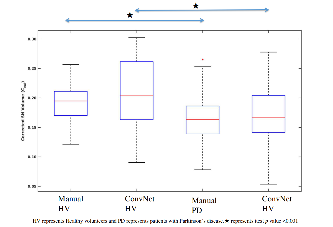
Figure 2: Box plot showing Corrected Substantia Nigra Pars Compacta (SNc) volume (Cvol) using ConvNet and manual segmentations.
