3207
Distinguishing malignant and benign lung lesions with multiparametric 18F-FDG PET/MRI: Comparison of metabolic parameters and IVIM parameters1Department of Medical Imaging, Henan Provincial People’s Hospital, People’s Hospital of Zhengzhou University, Zhengzhou, China, 2Department of Radiology, Zhengzhou University People’s Hospital & Henan Provincial People’s Hospital, Academy of Medical Sciences, Zhengzhou, China, 3Department of Radiology, Henan University People’s Hospital & Henan Provincial People’s Hospital, School of Basic Medicine. Department of Radiology, Zhengzhou University People’s Hospital & Henan Provincial People’s Hospital, Academy of Medical Sciences., Zhengzhou, China, 42258 Chengbei Road, Jiading District, Shanghai, China 201907, Shanghai, China
Synopsis
Distinguishing malignant from benign pulmonary lesions is critical. In the study, 18F-FDG PET/MRI were used to obtain metabolic parameters and IVIM diffusion parameters for differential diagnosis of benign and malignant lung lesions. Our results showed that multiparametric PET/MRI is helpful in the differentiation of pulmonary lesions. 18 FDG hybrid PET-MRI enables a robust differentiation of malignant and benign pulmonary lesions when several IVIM parameters and PET parameters are combined. Thus, PET-MRI may reduce some unnecessary lung biopsies.
Introduction
Lung cancer is the most common malignant tumor and its morbidity and mortality are increasing year by year. The differential diagnosis of benign and malignant pulmonary lesions is mainly based on morphological features, such as size, lobulation sign, spiculation sign, etc.; Computed tomography (CT) is commonly used to evaluate pulmonary lesions. Although many typical morphological features have been described for differential diagnosis, radiologists still have some challenges when using CT as a single imaging method for differential diagnosis of malignant and benign lung lesions, because there is a certain intersection between pulmonary benign and malignant lesions in CT findings. With the development of rapid imaging technology, PET/MRI has been used in clinic as the most advanced equipment in the imaging field, and has shown great application prospect in oncology [1,2]. Recently, although PET/MRI has been used in lung [3], its value in the differential diagnosis of benign and malignant pulmonary lesions remains to be further explored.Intravoxel incoherent motion (IVIM) model of DWI was first proposed by Le Bihan et al. [4,5,6], who concluded that it can allow separate evaluation of blood microcirculation and water-molecule diffusion. Recently, IVIM sequence has been successfully applied in the body, showing great potential in separate reflection of diffusion and perfusion without contrast agent injection [7,8,9]. With the development of rapid acquisition technology and artifact removal technology, IVIM sequence also has been successfully applied in the lung [10]. However, the value of IVIM model in detecting lung lesions has not been well investigated .
Thus, the aim of the study is to assess the value of multiparametric 18F-FDG PET/MRI in the differentiation of malignant and benign pulmonary lesions and compare the diagnostic efficiency of various quantitative parameters obtained from PET or IVIM .
Materials and Methods
This study was approved by the local institutional review board. Written informed consent was obtained from all the patients.Between June 2020 and November 2020, a total of 33 patients with pulmonary lesions (including 23 malignant and 10 benign pulmonary lesions). All the patients underwent preoperative PET/MRI by using a 3.0T PET/MR scanner (uPMR 790; UIH, Shanghai, China) with a 12 channels phased-array body coil. The PET tracer is 18F‑FDG with radiochemical purity >95%. All patients fasted for at least 6 hours and had a normal blood glucose level. The PET/MRI images were acquired 40-60 minutes of quiet rest after intravenous injection according to the standard dose of 0.15 mCi per kilogram weight. IVIM-DWI was set with the following parameters: TR= 1620 ms, TE= 69.6 ms. Thirteen different b-values were used: 0, 25, 50, 100, 150, 200, 400, 600, 800 and 1000 sec/mm2. Metabolic parameters including SUVmax, TLG and MTV were obtained from PET; Diffusion parameters including the pseudo-diffusion coefficient (Dp), true diffusion coefficient (Dt), perfusion fraction (F) and standard apparent diffusion cofficient (sADC) were calculated from IVIM DWI. All statistical analyses were performed using SPSS 25.0 and MedCalc12.0. The Mann-Whitney U test, Spearman rank correlation and receiver operating characteristic (ROC) analysis were performed for significant parameters to compare the diagnosis performance for detecting malignant and benign pulmonary lesions. P values less than 0.05 were considered significantly different.
Results
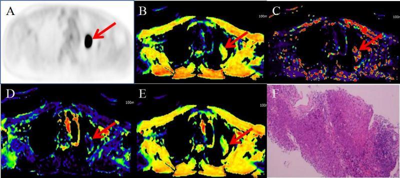
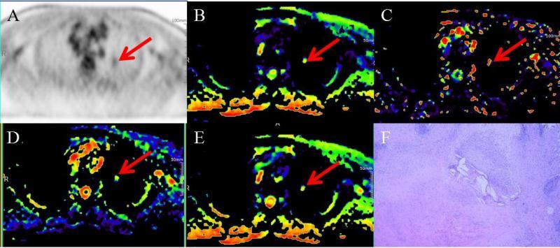
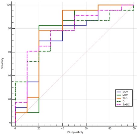
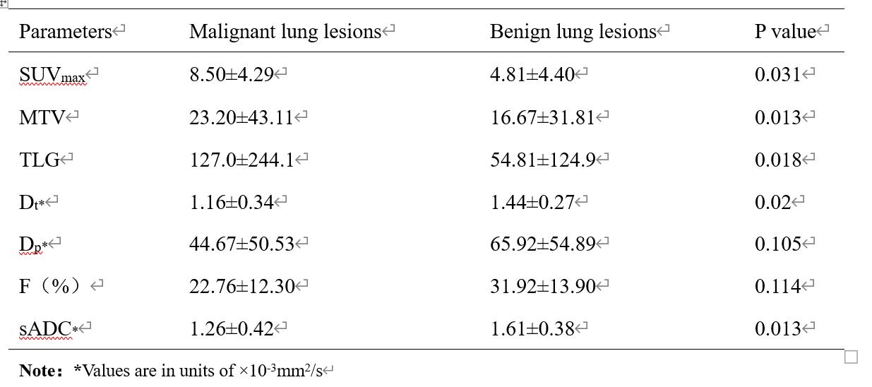
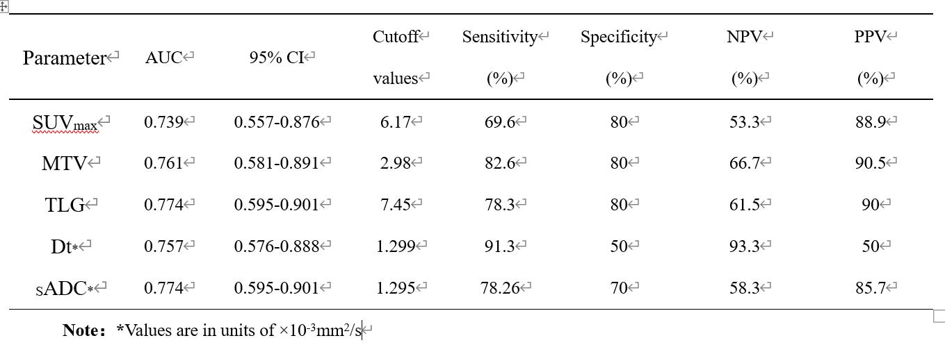
Dt and sADC values were lower (all P<0.05), while SUVmax, TLG and MTV were higher in malignant pulmonary lesions(Figure1) (all P<0.05) compared with benign pulmonary lesions (Figure2) (Table 1). Both Dp and F values showed no significant difference between malignant pulmonary lesions and benign pulmonary lesions (P>0.05).The area under the ROC curve (AUC) of SUVmax, TLG, MTV, Dt and sADC in discriminating malignant and benign pulmonary lesions are as follows: 0.739, 0774, 0.761, 0.757 and 0.774 (Figure1), but there were no significant differences among the diagnostic performance of each parameter(P>0.05) (Table 2). Furthermore, the AUC value of the combined parameters (SUVmax, TLG, MTV, Dt and sADC) in discriminating malignant and benign pulmonary lesions is 0.835 which is higher than that of any other single parameter.
In addition, there was a positive correlation between MTV and FTG (r = 0.934, P< 0.0001), between SUVmax and MTV (r=0.547, P < 0.05), between SUVmax and TLG (r= 0.758, P < 0.0001), between Dt and sADC (r=0.775, P < 0.0001). Additionally, there was a weakly negative correlation between Dt and SUVmax (r = -0.370, P < 0.05).
Discussion
Our results demonstrated that Dt and sADC were lower while SUVmax, TLG and MTV were higher in malignant lung lesions compared with benign lung lesions. However, both Dp and F showed no statistically significant difference between malignant and benign pulmonary lesions, implying that they might not be a potentially useful parameter for discriminating lung lesions. In addition, SUVmax, TLG, MTV, D and sADC all show moderate diagnostic ability and can be used as independently quantitative indexes and have potential value. Furthermore, for the differentiation of pulmonary lesions, the combination of SUVmax, TLG, MTV, D and sADC has higher diagnostic performance.Conclusion
SUVmax, TLG, MTV, Dt and sADC can be used as reliable independent indexes for distinguishing malignant and benign pulmonary lesions, and the combination of several IVIM parameters and PET parameters could improve the differentiation of malignant and benign pulmonary lesions.Acknowledgements
This research was supported by National Key R&D Program of China (2017YFE0103600), National Natural Science Foundation of China (81720108021), Zhongyuan Thousand Talents Plan Project(ZYQR201810117), Zhengzhou Collaborative Innovation Major Project (20XTZX05015)References
1. Schulz V, Torres-Espallardo I, Renisch S, et al. Automatic, three-segment, MR-based attenuation correction for wholebody PET/MR data. Eur J Nucl Med Mol Imaging. 2011; 38(1): 138-152.
2. Sawicki LM, Grueneisen J, Buchbender C, et al. Comparative performance of 18F-FDG PET-MRI and 18FFDG PET-CT in detection and characterization of pulmonary lesions in 121 oncologic patients. J Nucl Med. 2016; 57(4): 582-586
3. Chandarana H, Heacock L, Rakheja R, et al. Pulmonary nodules in patients with primary malignancy: comparison of hybrid PET/MR and PET/CT imaging. Radiology.2013;268(3): 874-881.
4. Le Bihan D, Breton E, Lallemand D, et al. MR imaging of intravoxel incoherent motions: application to diffusion and perfusion in neurologic disorders. Radiology.1986;161(2): 401-407.
5. Le Bihan D, Breton E, Lallemand D, et al. Separation of diffusion and perfusion in intravoxel incoherent motion MR imaging. Radiology. 1988;168(2):497-505.
6. Y amada I, Aung W, Himeno Y, et al. Diffusion coefficients in abdominal organs and hepatic lesions: evaluation with intravoxel incoherent motion echo-planar MR imaging. Radiology. 1999;210(3):617-623
7. Franca M, Marti-Bonmati L, Alberich-Bayarri A, et al. Evaluation of fibrosis and inflammation in diffuse liver diseases using intravoxel incoherent motion diffusion-weighted MR imaging. Abdom Radiol (NY).2017;42(2):468–477.
8. Winfield JM, Desouza NM, Priest AN, et al. Modelling DW-MRI data from primary and metastatic ovarian tumours. Eur Radiol. 2015; 25(7):2033-2040.
9. Koh DM, Collins DJ, Orton MR. Intravoxel incoherent motion in body diffusion-weighted MRI: reality and challenges. AJR Am J Roentgenol. 2011; 196(6): 1351–1361
10. Wang LL; Lin J;Liu K et al. Intravoxel incoherent motion diffusion-weighted MR imaging in differentiation of lung cancer from obstructive lung consolidation: comparison and correlation with pharmacokinetic analysis from dynamic contrast-enhanced MR imaging. Eur Radiol.2014; 24(8):1914-22
Figures