3201
The assessment of cartilage endplate and its relationship with the corresponding disc degeneration using UTE imaging1The First Affiliated Hospital of Nanjing Medical University, Nanjing, China, 2GE Healthcare, MR Research China, Beijing, P.R. China, Beijing, China
Synopsis
In this study, we aimed to investigate the feasibility of ultra-short echo time (UTE) imaging for assessing the cartilage endplate (CEP) damage and evaluate the relationship between the grading of CEP and lumbar disc degeneration. UTE images with short and long TEs were performed on patients with low back pain. The resultant subtracted UTE images between short and long TEs showed well structural CEP clearly. Strong correlation was observed between CEP degree based on UTE imaging and lumbar intervertebral disc degeneration. With these findings, UTE imaging might thus be considered an effective tool to assess the CEP damage in clinic.
Introduction
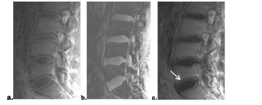
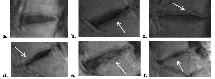
Eighty-four percent of people have low back pain in their lifetime. The cartilage endplate (CEP) lesion is regarded as one important reason for this disease1. Using conventional T1-weighted MRI, S. Rajasekarank et al. classified endplate as six levels (Fig.1). With this criterion, a so-called Total Endplate damage Score (TEPS) is calculated to help for clinic treatment2. However, due to short T2 relaxation time, CEP Imaging using conventional MRI usually suffers low signal and is challenged for well structural depiction3.Ultra-short Echo Time (UTE) MR imaging, as a relatively novel technique for high signal-to-noise ratio (SNR), has been applied in CEP4. With flexible selections of short or long TEs, UTE images dominated with bound & free water protons or free water protons only can be acquired respectively5. Bound water protons dominated UTE images can thus be easily obtained by subtracting UTE images from short to long TEs, showing CEP with well depicted structures4,6(Fig.2). While promising anatomical imaging was obtained, no UTE relevant study has however, been performed to further evaluate TEPS for patients with low back pain. This is actually crucial as CEP damage is assumed to be more accurately evaluated with better anatomical imaging.
Therefore, this study aimed to explore the feasibility of UTE imaging in accurate assessment of the extent of CEP damage. Additionally, the resultant CEP grading was further correlated with lumbar disc degeneration evaluated by clinical Pfirmann score system7.
Materials and method
Subjects:33 patients with low back pain (mean: 47±19 years) were recruited in this study. These patients had no history of spinal surgery or radiotherapy, and were also excluded with spinal deformity, trauma, benign and malignant vertebral tumors, hematologic diseases and other systemic diseases.
MRI experiment:
All experiments were performed on a 3T MRI (Discovery 750w, GE Healthcare, USA) equipped with Large Flexible Array coil.
For double-TE UTE imaging, L1-S2 with field of view of 36.0mm×36.0mm were measured at sagittal view. Other scan parameters were of repetition-time =12.5ms, short and long TEs=0.03ms and 6ms, slice thickness=4.0 mm, number of slices=20 and bandwidth=83.33Mhz. A total scan time was 2 minutes 48s.
Data analysis:
All acquired UTE images were analyzed in ADW4.6 workstation (GE Medical Systems).
Bound water dominated UTE images of CEP were calculated with a simple subtraction of UTE images from short to long TEs. To evaluate the image quality, SNR and contrast-to-noise ratio (CNR) were calculated for the resultant subtracted UTE images:
$$SNRx =|Sx-Sbackground|/s$$
and
$$CNR(x-y)=|Sx-Sy|/s$$
where Sx, Sy and Sbackground represent the mean signal intensities of ROIs x for the most clear area of CEP, y for the maximum contrast region and background, respectively, and s is the standard deviation (SD) of the background noise.
Two senior radiologists were independently employed to assess the CEP damage and lumbar intervertebral discs’ (IVD) degeneration based on the subtracted UTE images and T2-weight images. For CEP assessment, endplate classification criterion2(Fig.1), defined to evaluate the severity of endplate damage in six types, was applied. For IVD, Pfirrman grading was used to evaluate the degeneration severity of IVD in five types7.
Statistical analysis:
All statistical analyses were performed using SPSS software. Kappa test was used to evaluate the inter-observer agreement in assessing the degree of CEP defect and lumbar disc degeneration between two radiologists. Kendall's TAU-B test was used to analyze the correlation between CEP injury and lumbar disc degeneration. P<0.05 was set the threshold of statistical significance.
Results
Well structural CEPs were clearly shown in subtracted UTE images of CEPS (Fig.2c). Additionally, the image quality was also confirmed by high SNR and CNR (mean SNR: 33.06±2.92; mean CNR: 9.4±2.08) .Using Cohen's Kappa test, excellent inter-observer agreement between two radiologists was confirmed by high coefficient of 0.922 (95%CI:0.925~1.059) and 0.769 (95%CI:0.882~1.102) in assessing the CEP damage and IVD degeneration (both P<0.001), respectively.
Ensured by reliable evaluation, 77 intervertebral discs were selected from 33 patients. Six grades were divided according to the integrity of CEP (Fig.3). Fifteen of the CEPs were type I, fifteen were type II, thirteen were type III, twelve were type IV, ten were type V, twelve were type VI. The corresponding discs degeneration was divided into five grades according to Pfirmann grading.
Kendall's TAU-B analysis was also applied to evaluate the relationship between the CEP grades and the lumbar disc degeneration evaluated by Pfirmann score system. High coefficient of 0.818 was obtained, revealing a significantly strong correlation (P<0.001).
Discussion and conclusion
In this study, we investigated the feasibility of UTE imaging in assessing the damage of CEPs for patients with low back pain. Using subtracted UTE images from short to long TEs, well structural CEP images with high image quality were obtained and classified into 6 grades according to endplate classification criterion. The TEPs could thus be calculated for further clinical treatment. In addition, high positive correlation was found between UTE based CEP grading and IVD grading evaluated with clinical Pfirrman scoring, indicating that the increase of the CEP degree might induce more serious the disc degeneration.In conclusion, with these findings, UTE imaging might be considered an effective tool in assessing CEP damage in clinic.
Acknowledgements
Thank the orthopedic doctor for his case and cooperation of enrolled patients.References
1. Adams MA, Roughley PJ. What is intervertebral disc degeneration, and what causes it? Spine. 2006;31(18):2151-2161.
2. Rajasekaran S, Venkatadass K, Naresh Babu J, et al. Pharmacological enhancement of disc diffusion and differentiation of healthy, ageing and degenerated discs: Results from in-vivo serial post-contrast MRI studies in 365 human lumbar discs. Eur Spine J. 2008;17(5):626-643.
3. Bae WC, Biswas R, Chen K, et al. UTE MRI of the Osteochondral Junction. Current radiology reports. 2014; 2(2):35.
4. Berg-Johansen B, Han M, Fields AJ, et al. Cartilage Endplate thickness variation measured by Ultrashort Echo-Time MRI is associated with adjacent disc degeneration. Spine. 2018;43(10):592-600.
5. Du J, Bydder M, Takahashi AM, et al. Short T2 contrast with three-dimensional ultrashort echo time imaging. Magn Reson Imaging. 2011;29(4):470-482.
6. Kim YJ, Cha JG, Shin YS, et al. 3D Ultrashort TE MRI for evaluation of cartilaginous endplate of cervical disk in vivo: Feasibility and correlation with disk degeneration in T2-weighted spin-echo sequence. AJR Am J Roentgenol. 2018;210(5):1131-1140.
7. Pfirrmann CW, Metzdorf A, Zanetti M, et al. Magnetic resonance classification of lumbar intervertebral disc degeneration. Spine. 2001;26(17):1873-1878.
Figures