3196
Multimodal MRI evaluation of paraspinal muscles and TNF- α in rats with discogenic low back pain after rehabilitation training1Medical imaging department, The First Affiliated Hospital of Kunming Medical University, Kunming, China, 2GE Healthcare,MR Research China, Beijing, China
Synopsis
Based on functional MRI, the current study aims to explore the pathological morphology, fat infiltration and the expression of TNF-α in paraspinal muscles of discogenic low back pain(DLBP). And to further explore the relation of rehabilitation exercise on paraspinal muscles and intervertebral discs in rats. It is concluded that rehabilitation exercise can reduce the content of TNF-α in serum and paraspinal muscles, relieve the symptoms of DLBP in rats, and improve paraspinal muscle dysfunction.
Introduction/ Purpose
Low back pain (LBP) has become the leading cause of disability worldwide, of which DLBP accounts for 39% [1, 2]. The clinical manifestation of DLBP is lack of specificity, and the exact pathogenesis remains unclear. Although the prevalence rate is high, the effective treatment strategies available are very limited. Exercise therapy is one of the most commonly used and effective methods to treat DLBP, which is also an important means of modern clinical rehabilitation, but its mechanism is still unclear [3]. Therefore, based on functional MRI, this study explores the possible pathogenesis of DLBP and the mechanism of rehabilitation exercise on intervertebral discs and paraspinal muscles in rats.Material and Methods
Study design1. Under the guidance of X-ray fluoroscopy, the lumbar intervertebral discs of L4/L5 and L5/L6 of SD rats were punctured laterally and posteriorly with by 25G needle, and 2.5μl of PBS was injected into the nucleus pulposus to establish the model of DLBP in rats.
2. Swimming rehabilitation exercise was started 1 day after operation for 5 days, 30 minutes a day. Every day each rat swims in a cylindrical container with a diameter of 17cm, a height of 40cm and a depth of 30cm. The water temperature is between 34℃ and 36℃.
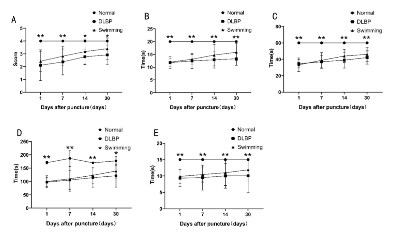
3. Behavioral evaluation was performed at 1 day, 1 week, 2 weeks and 1 month after operation. Gait test was used to evaluate the gait and hindlimb use, hot plate test to evaluate the response threshold of rats to thermal stimulation, acetone test to evaluate the response threshold of rats to cold stimulation, tail suspension test to evaluate the lumbar muscle strength of rats, and grip strength test to evaluate the waist and limbs strength of rats.
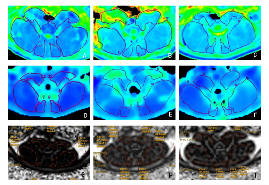
4. After operation, the paraspinal muscle dysfunction and fat infiltration of rats were evaluated by multimodal magnetic resonance imaging, including T2WI, T2-mapping, BOLD and IDEAL-IQ sequences.
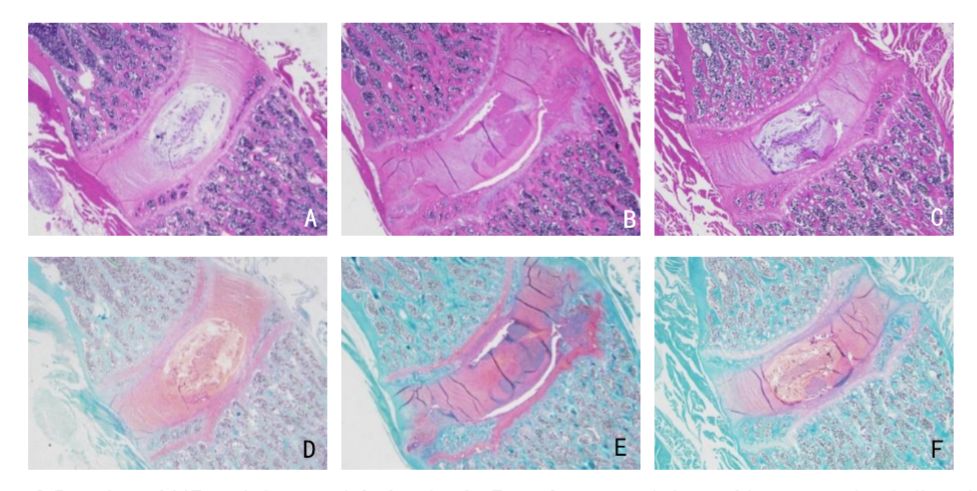
5. After operation, the structural degeneration of nucleus pulposus and annulus fibrosus and the changes of extracellular matrix were evaluated by HE staining and Safranin O-Fast Green staining.
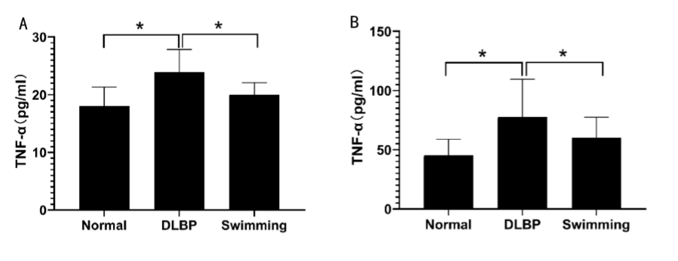
6. Elisa test was used to evaluate the expression of TNF-α in paraspinal muscle and serum after rehabilitation exercise.
MRI and Imaging processing
All subjects underwent MR scanning after one-month operation on a 3.0 T MRI scanner (Discovery MR 750W, GE Healthcare, Waukesha, USA) using 3.0T 16-channel longitudinal mouse coil. The following three imaging protocols were performed: A) T2 mapping: TE=8.6-69.1ms, TR=853ms, FOV=8cm×8cm, Bandwidth=41.67Hz, NEX=1, slice thickness=2.9mm, matrix=256×256; B) BOLD: TE=2.3-34.3ms, TR=100ms, FOV=8cm×8cm, Bandwidth=31.25Hz, NEX=2, slice thickness=2.9mm,matrix=192×192; C)IDEAL IQ: TE=1.8-7.3ms, TR=14.4ms, FOV=15cm×15cm, Bandwidth=83.33Hz, NEX=2, slice thickness=2.0mm, matrix=178×178.Other imaging parameters included: FOV=10×10cm, matrix=256×256, slice thickness=1.2mm. The total scan time was 39.48mins.
Statistical methods
All analyses were performed using the statistical software SPSS for Windows version 22.0 (IBM SPSS Inc., IL, USA). Kruskal-Wallis test were used to evaluate the relationship between behavioral evaluation and imaging evaluation of three groups one month after operation. One-way ANOVA (analysis of variance) were used to compare the differences of TNF-α content in muscle and serum of three groups of rats one month after operation.
Results
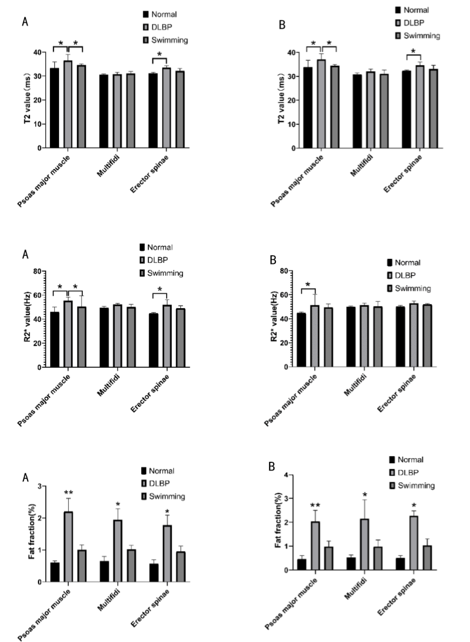
1.The behavior test showed that the gait score of swimming group was higher than that of DLBP group (P<0.05). Compared with the DLBP group, the hot and cold stimulation threshold of the swimming group was higher. Tail suspension test showed that the swimming group spent more time bending and struggling than the DLBP group. The grip strength test found that the waist and limbs were stronger in the swimming group.2.The imaging results showed that there were significant differences in T2 value, R2* value and fat fraction among the three groups (P<0.05). The pseudo-color map showed that the values of T2, R2 * and fat fraction in the swimming group were lower than those in the DLBP group.
3. The results of histological staining showed that the shape of intervertebral disc was irregular, nucleus pulposus pyknosis, fibrous annulus disordered and irregular edge of cartilage in DLBP group, and the shape of intervertebral disc was significantly improved in swimming group.
4. Elisa test showed that the expression of TNF-α in serum and paraspinal muscles increased in DLBP group, but decreased significantly in swimming group(P<0.05).
Discussion and Conclusion
The results of this study showed that the behavioral score of DLBP rats decreased, indicating a decrease in pain threshold and lumbar function. Multimodal MRI showed fat infiltration in paraspinal muscle, irregular shape of intervertebral disc could be seen by histological staining, and the content of TNF-α in paraspinal muscle and serum was increased by Elisa test. It is suggested that intervertebral disc degeneration will lead to the increase of TNF-α, paraspinal muscle fat infiltration and spinal instability, thus aggravating intervertebral disc degeneration. Rehabilitation exercise is the first choice for the treatment of DLBP, and it has been reported that rehabilitation training can improve the function of paraspinal muscles [4] by reducing TNF-α [5]. The results showed that after rehabilitation training, the behavioral score of rats increased, the fat infiltration of paraspinal muscle improved, and the content of TNF-α in serum and paraspinal muscle decreased, suggesting that rehabilitation training can improve fat infiltration in paraspinal muscles by reducing TNF-α content, thus relieving low back pain.Acknowledgements
No acknowledgment found.References
[1] Lu Y, Guzman JZ, Purmessur D, et al. Nonoperative management of discogenic back pain: a systematic review. Spine (Phila Pa 1976). 2014. 39(16): 1314-24.
[2] Maher C, Underwood M, Buchbinder R. Non-specific low back pain. Lancet. 2017. 389(10070): 736-747. [3] Bernstein IA, Malik Q, Carville S, Ward S. Low back pain and sciatica: summary of NICE guidance. BMJ. 2017. 356: i6748.
[4] Wang CK, Fang YD, Lin LC, et al. Magnetic Resonance Elastography in the Assessment of Acute Effects of Kinesio Taping on Lumbar Paraspinal Muscles. J Magn Reson Imaging. 2019. 49(4): 1039-1045.
[5] Al-Obaidi S, Mahmoud F. Immune responses following McKenzie lumbar spine exercise in individuals with acute low back pain: a preliminary study. Acta Med Acad. 2014. 43(1): 19-29.
Figures