3193
Evaluation of Lumbar Disc Degeneration Using Ultrashort Echo Time Magnetization Transfer (UTE-MT) Imaging in Cartilaginous Endplate1Department of Radiology, The Fifth Affiliated Hospital of Sun Yat-Sen University, Zhuhai, China, 2Department of Radiology, University of California, San Diego, CA, United States, 3Department of Orthopedics, The Fifth Affiliated Hospital of Sun Yat-Sen University, Zhuhai, China, 4MR Research, GE Healthcare, Guangzhou, China
Synopsis
Ultrashort echo time magnetization transfer (UTE-MT) sequence can quantify magnetization transfer ratio (MTR) values of short T2 tissues, which has potential to evaluate early disc degeneration. The current study aims to assess the feasibility of UTE-MT quantification of cartilaginous endplate for evaluation of early disc degeneration. In addition, the latent associations between the UTE-MTR and grade of disc degeneration, age, gender, the oswestry disability index (ODI) were evaluated, respectively. Our results demonstrated that the UTE-MT sequence is feasible to quantify cartilaginous endplate in lumbar spine. UTE-MTR may be a potentially useful biomarker in the assessment of early lumbar disc degeneration.
Introduction
Degeneration or calcification of cartilaginous endplate may be highly related to the early changes of intervertebral disc. However, the T2 of cartilaginous endplate is relatively short, which cannot be effectively imaged with clinical sequences.[1] Ultrashort echo time magnetization transfer (UTE-MT) sequence with TEs less than 100 μs can be utilized to image and quantify both short and long T2 tissues. The derived biomarker of magnetization transfer ratio (MTR) [2] may be potentially useful for early disc degeneration assessment. In this study, we utilized UTE-MT sequence to investigate the correlations between UTE-MTR of cartilaginous endplate and disc degeneration, age, gender, ODI, respectively, in disc degeneration patients.Methods
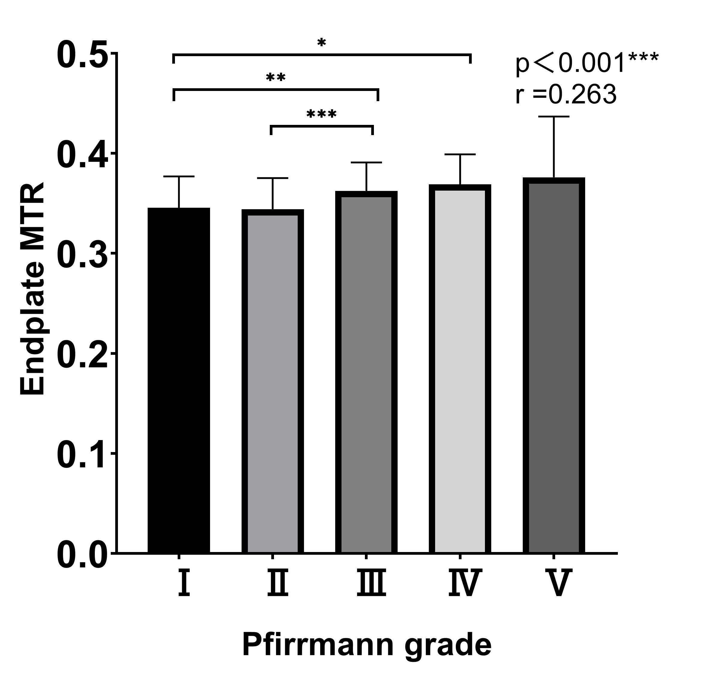
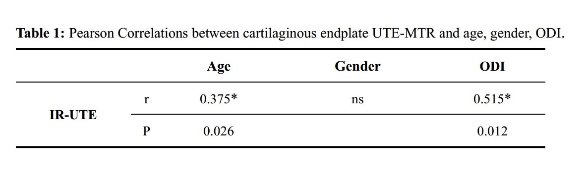
A total of 62 disc degeneration patients (age 43 ± 17 years, age range 30-78 years) were recruited and underwent UTE-MT sequence of lumbar on a 3.0T MRI scanner (Signa, Pioneer, GE Healthcare). A Fermi pulse was employed to generate the MT contrast in UTE-MT sequence with duration of 8ms and bandwidth of 160Hz. The frequency offset of this MT pulse was 1500 Hz. The UTE-MT sequence was scanned twice with flip angle of 750° for MT-On and 0° for MT-Off. Other UTE-MT sequence parameters were as follows: TR = 100 ms, TE = 0.032 ms, excitation flip angle = 5°, number of spokes per-TR = 5, FOV = 28cm × 28cm, matrix = 140 × 140, slice thickness = 3.6mm, and slice number = 16, oversampling factor = 1.2, and scan time = 3min. The ROI of the cartilaginous endplate was manually delineated by a radiologist with 10 years of experience. Mean cartilaginous endplate UTE-MTR value was computed for each disc by averaging the UTE-MTR measurements of adjacent upper and lower cartilaginous endplates. The grade of disc degeneration level was assessed using the Pfirrmann scoring system. Pearson correlation analysis was performed to calculate the correlations between cartilaginous endplate UTE-MTR and grade of disc degeneration, age, gender, ODI, respectively. A One way ANOVA test was used to compare cartilaginous endplate UTE-MTR between different grades of disc degeneration. A value of P < 0.05 was considered as a statistically significant.Results
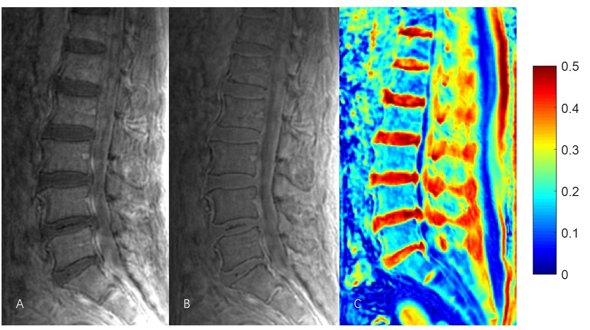
Figure 1 shows the representative lumbar UTE-MT images acquired from a 67-year-old male lumbar disc degeneration volunteer. The cartilaginous endplate UTE-MTR showed a strong positive correlation with Pfirrmann grade of disc (r= 0.263, P < 0.001) (Figure 2), cartilaginous endplate UTE-MTR increased with Pfirrmann grade of disc. In addition, our results demonstrated that the cartilaginous endplate UTE-MTR was negatively correlated with age (r = 0.375*, P = 0.026) and ODI (r = 0.515, P = 0.012), while had no association with gender. The results were summarized in table 1.Discussion and Conclusion
To our best knowledge, this is the first prospective study of applying UTE-MT in cartilaginous endplate to assess disc degeneration. Our findings suggested that deficits in CEP composition, as indicated by high UTE-MTR, associate with more severe disc degeneration in patients. The high UTE-MTR of cartilaginous endplate may be related to the decrease of proteoglycan and collagen in cartilaginous endplate.[3] Overall, it is supposed that UTE-MTR of cartilaginous endplate may be a useful biomarker to predict early disc degeneration.Acknowledgements
No acknowledgement found.References
[1] Q. Song, X. Liu, D. J. Chen, Q. Lai, B. Tang, B. Zhang, M. Dai and Z. Wan, "Evaluation of MRI and CT parameters to analyze the correlation between disc and facet joint degeneration in the lumbar three-joint complex," Medicine (Baltimore), vol. 98, no. 40, pp. e17336, 2019.
[2] Y. J. Ma, E. Y. Chang, M. Carl and J. Du, "Quantitative magnetization transfer ultrashort echo time imaging using a time-efficient 3D multispoke Cones sequence," Magn Reson Med, vol. 79, no. 2, pp. 692-700, 2018.
[3] R. J. Moore, "The vertebral end-plate: what do we know?," Eur Spine J, vol. 9, no. 2, pp. 92-96, 2000.
Figures