3191
Effects of different acceleration factors on lumbar body fat quantification using 3D mDixon Quant technique
Yu Song1, Qingwei qing Song2, Yingkun Guo1, Gang Ning1, and Xuesheng Li1
1Department of Radiology, West China Second University Hospital, Sichuan University, Chengdu, China, 2Department of Radiology, the First Affiliated Hospital of Dalian Medical University, Dalian, China
1Department of Radiology, West China Second University Hospital, Sichuan University, Chengdu, China, 2Department of Radiology, the First Affiliated Hospital of Dalian Medical University, Dalian, China
Synopsis
Compressed SENSE (CS) is a cutting-edge signal acquisition and processing innovation technology based on applied mathematics. By directly collecting the compressed signal, the amount of sampled data is greatly reduced, and the optimal acceleration factor can be passed. The realization of the method can shorten the clinical magnetic resonance scanning time while ensuring the spatial resolution.
Synopsis
Compressed SENSE (CS) is a cutting-edge signal acquisition and processing innovation technology based on applied mathematics. By directly collecting the compressed signal, the amount of sampled data is greatly reduced, and the optimal acceleration factor can be passed. The realization of the method can shorten the clinical magnetic resonance scanning time while ensuring the spatial resolution.Purpose
To investigate the effects of different acceleration factors (AF) on lumbar vertebra fat quantification using 3D mDixon Quant sequence accelerated by sensitivity coding (SENSE) and Compressed SENSE(CS-SENSE).Materials and Methods
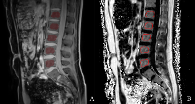
From January to February 2020, 96 healthy volunteers were recruited, including 45 males and 51 females, from 16 to 79 years old, with an average age of 43.85 ± 17.98 years. All volunteers were imaged at 3.0T (Ingenia CX 3;Philips Healthcare, Best, the Netherlands) with a fat quantification sequence (3D mDixon Quant) for the entire lumbar spine, and the sequence was accelerated at different folds with either SENSE (acceleration factor S = 2, 4) or CS-SENSE (acceleration factor CS =2,3,4,5,6,7,8). FF, signal-noise-ratio (SNR), and contrast-noise-ration (CNR) were measured for the L1-L5 vertebra by two radiologists independently with a double-blind method, measurement carried out on a IntelliSpace Portal workstation (ISP version 7.0; Philips Healthcare, Best, the Netherlands)(Figure 1). The measured parameters were compared among different AFs. The data were analyzed by SPSS 19.0 statistical software. The intra-class correlation coefficients (ICC) were used to check the consistency of the measurement between the two observers. The non-parametric Friedman test was used to analyze the differences in FF, SNR and CNR measured at different AFs. P <0.05 was considered statistically significant.Results
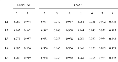
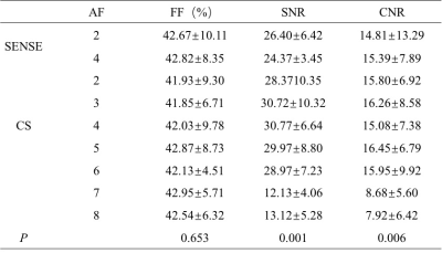
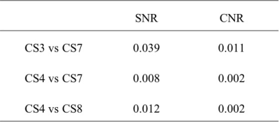
The measurement results of the two observers showed good agreement (ICC value>0.75)(Table 1). There was no statistically significant difference in the FF values measured by 3D mDixon Quant sequence with different AF (P=0.653), but there were statistically significant differences in SNR and CNR with different AF (P=0.001, 0.006)(Table 2). Among them, there were statistically significant differences in SNR and CNR between CS3 and CS7 groups, CS4 and CS7 groups, and CS4 and CS8 groups (P<0.05)(Table 3). The scanning time of CS6 was 60.66% shorter than that of SENSE 2, but there were no statistically significant differences in FF values, SNR and CNR values compared with those of other groups (S2, S4, CS2, CS3, CS4 and CS5) (P>0.05).Discussion
Compressed SENSE technology is a cutting-edge signal acquisition and processing innovation technology based on applied mathematics[1]. It uses digital sparse sampling to obtain a small amount of sampled data and ensure the fidelity of the image. The signals collected in K space are converted by Fourier transform and wavelet transform After conversion to Hilbert space, fine discrete noise reduction can be achieved[2]. Compressed SENSE technology greatly reduces the amount of sampled data by directly collecting the compressed signal, and achieves a significant reduction in magnetic resonance scanning time[3-4]. However, studies have shown that the larger the acceleration factor, the shorter the corresponding scanning time, but at the same time the image quality will be affected to a certain extent, the image signal-to-noise ratio is reduced, and curl artifacts are likely to occur[5]. Therefore, it is very important to choose the best factor when scanning different clinical sequences in different parts.Conclusion
The 3D mDixon Quant sequence combined with SENSE and CS technology is reliable for assessing the lumbar vertebral body fat content. When the AF of CS was selected to 6, the image quality and the measurement accuracy were maintained while the imaging time dramatically reduced.Acknowledgements
No acknowledgement found.References
No reference found.Figures

Figure.1 Male, 24 years old, BMI= 21.47kg /m2, Fig. 4A and Fig. 4B are 3D mDixon Quant water image and FF image, respectively

Table 1 Consistency test of results measured by two observers (ICC value)

Table 2 The difference of lumbar vertebrae fat fraction(FF) and image quality in different AF

Table 3 Pairwise comparison in different AF groups with different image quality (P value)