3188
A comparative study of R2*, UTE-T2* and T1rho mapping for evaluation of degenerative alterations in human intervertebral discs
LiLan Wu1, JianJun Zhou2, and Pu-Yeh Wu3
1Department of Radiology, Fudan University Affiliated Zhongshan Hospital Xiamen Branch, Xiamen, China, 2Department of Radiology, Fudan University Affiliated Zhongshan Hospital, Shanghai, China, 3GE Healthcare, Beijing, China
1Department of Radiology, Fudan University Affiliated Zhongshan Hospital Xiamen Branch, Xiamen, China, 2Department of Radiology, Fudan University Affiliated Zhongshan Hospital, Shanghai, China, 3GE Healthcare, Beijing, China
Synopsis
This study aims to investigate whether human lumbar IDD can be detected using R2*, UTE-T2* and T1rho mapping. We systematically compared the efficacy of R2*, UTE-T2* and T1rho values in the diagnosis of early IDD. Specifically, we found that T1rho value is superior to UTE-T2* and R2* values for diagnosis of the early IVD, while UTE-T2* value is optimal for diagnosis of advanced IVD. Overall, we concluded that R2* and UTE-T2* mapping provides another promising method for quantitatively evaluated lumbar IDD, and T1rho mapping can be considered an effective tool for distinguishing IDD at earlier stage of the degenerative process
Introduction
Low back pain (LBP) is one of the main causes leading to disability worldwide, resulting in a great burden on global health care system [1, 2]. A number of studies have reported that intervertebral disc degeneration (IDD) is a significant contributor of nonspecific LBP, with a lifetime prevalence of over 80% [3, 4]. Early stages of IDD are mainly in the form of biochemical changes, including glycosaminoglycan decreasing. It will lead to a decrease of hydrostatic pressure, resulting in nucleus pulposus (NP) dehydration as well as loss of structural and mechanical properties of intervertebral discs (IVDs). Detecting early changes in IDD is important for developing preventative strategies or reestablishing degenerated IVDs, such as nucleus replacement, cell therapy, and growth factor therapy [5]. Conventional T2 mapping is sensitive to changes of tissues with relatively long T2 relaxations, but limited in identifying early biochemical changes of IVDs [5]. Ultrashort echo time (UTE) imaging could catch very short T2/T2* signals (< 10 ms) [6-9], and has been confirmed to be sensitive to deep tissue matrix changes of subtle and preclinical degeneration [10]. Additionally, previous studies showed that T1rho mapping is a promising technique for diagnosis of biochemical degeneration [11-13]. This preliminary study aims to assess whether human lumbar IDD can be detected using these quantitative approaches, including regular R2* mapping, UTE-T2* mapping and T1rho mapping. Furthermore, we systematically compared the efficacy of R2*, UTE-T2* and T1rho values in the diagnosis of early IDD.Materials and Methods
Seventy-six patients with nonspecific LBP were enrolled in this study. Subjects were divided into three age groups: 19-39 y, 40-60 y, and 60-85 y. All data were acquired on a 3.0T MRI scanner (Discovery MR750w, GE Healthcare, Milwaukee, WI). Patients underwent MRI examinations including: (1) sagittal T2WI(2) R2* mapping (3) UTE-T2* mapping; (4) T1rho mapping. T2WI were used for Modified 5-level Pfirrmann grading. Disc degenerations were also divided into three categories: normal (Pfirrmann grade I), early stage degeneration (Pfirrmann grade II-III), and advanced stage degeneration (Pfirrmann grade V). R2*, UTE-T2*, and T1rho maps were calculated by mono-exponential fitting using AW4.6 GE Workstation or a custom code in MATLAB (MathWorks, Natick, MA). Five circular regions of interest (ROIs) with equal size were manually drawn on T2WI images from anterior to posterior (Figure 1), including the anterior annulus fibrosus (AAF; ROI 1), the nucleus pulposus (NP; ROI 3), and posterior annulus fibrosus (PAF; ROI 5). The mean value of ROI 1-5 was treated as the averaged measurement of the whole disk. Statistical analysis was conducted using SPSS 22.0 software (IBM, Armonk, NY). Correlations of quantitative values with Pfirrmann grades and age were analyzed using Spearman's rank correlation. Receiver operating characteristic (ROC) analysis was performed and area under the curve (AUC), sensitivity and specificity were obtained to assess the diagnostic efficacy of each quantitative parameter in differentiating normal IVDs from early stage degeneration, and early stage degeneration from advanced stage degeneration. P value less than 0.05 was considered statistically significant.Results
We found that R2*, UTE-T2* and T1rho values in NP showed moderate correlations with Pfirrmann grade (r = -0.419, 0.480, -0.614, respectively; P < 0.001). No significant correlation was observed in R2* and UTE-T2* values in AAF with Pfirrmann grade. R2*, UTE-T2* and T1rho values in PAF showed low correlations with Pfirrmann grade (r = -0.210、0.218、-0.398, respectively; P < 0.001). The AUC values of the metrics for differentiating the normal and early stage degeneration IVDs ranked as follows: T1rho > UTE-T2* > R2*. For differentiating the early and advanced stage degeneration IVDs, the rank of the diagnostic efficacy was UTE-T2* > R2* > T1rho. Comparing the quantitative parameters in different regions, diagnostic performance of NP was the highest, and that of AAF was the lowest.Conclusion
The current study demonstrated the feasibility of quantitative evaluation of human IDD in vivo by R2*, UTE-T2* and T1rho mapping. We found that T1rho value is superior to UTE-T2* and R2* values for differential diagnosis of the early IVD, while UTE-T2* value is optimal for differential diagnosis of advanced IVD. Furthermore, the diagnostic performance of these quantitative parameters was highest in NP. Overall, we concluded that R2* and UTE-T2* mapping provides another promising method for quantitatively evaluated lumbar IDD, and T1rho mapping can be considered an effective tool for distinguishing IDD at the earlier stage of the degenerative process.Acknowledgements
References
1. BARDIN L D, KING P, MAHER C G. Diagnostic triage for low back pain: a practical approach for primary care[J]. Med J Aust, 2017,206(6): 268-273. 2. OGON I, TAKEBAYASHI T, TAKASHIMA H, et al. Imaging diagnosis for intervertebral disc[J]. JOR SPINE, 2020,3(1). 3. LIAO Z, LUO R, LI G, et al. Exosomes from mesenchymal stem cells modulate endoplasmic reticulum stress to protect against nucleus pulposus cell death and ameliorate intervertebral disc degeneration in vivo[J]. Theranostics, 2019,9(14): 4084-4100. 4. BASHKUEV M, REITMAIER S, SCHMIDT H. Relationship between intervertebral disc and facet joint degeneration: A probabilistic finite element model study[J]. J Biomech, 2020,102: 109518. 5. BERG-JOHANSEN B, HAN M, FIELDS A J, et al. Cartilage Endplate Thickness Variation Measured by Ultrashort Echo-Time MRI Is Associated With Adjacent Disc Degeneration[J]. SPINE, 2018,43(10): E592-E600. 6. CHANG E Y, Du J, BAE W C, et al. Qualitative and Quantitative Ultrashort Echo Time Imaging of Musculoskeletal Tissues[J]. Semin Musculoskelet Radiol, 2015,19(4): 375-386. 7. Du J, TAKAHASHI A M, CHUNG C B. Ultrashort TE spectroscopic imaging (UTESI): application to the imaging of short T2 relaxation tissues in the musculoskeletal system[J]. J Magn Reson Imaging, 2009,29(2): 412-421. 8. CHANG E Y, DU J, CHUNG C B. UTE imaging in the musculoskeletal system[J]. Journal of Magnetic Resonance Imaging, 2015,41(4): 870-883. 9. QIAO Y, TAO H, MA K, et al. UTE-T2* Analysis of Diseased and Healthy Achilles Tendons and Correlation with Clinical Score: An In Vivo Preliminary Study[J]. BioMed Research International, 2017,2017: 1-5. 10. CHU C R, WILLIAMS A A, WEST R V, et al. Quantitative Magnetic Resonance Imaging UTE-T2* Mapping of Cartilage and Meniscus Healing After Anatomic Anterior Cruciate Ligament Reconstruction[J]. Am J Sports Med, 2014,42(8): 1847-1856. 11. WANG Y J, ZHAO F, GRIFFITH J F, et al. T1rho and T2 relaxation times for lumbar disc degeneration: an in vivo comparative study at 3.0-Tesla MRI[J]. European Radiology, 2013,23(1): 228-234. 12. PAUL C P L, SMIT T H, de GRAAF M, et al. Quantitative MRI in early intervertebral disc degeneration: T1rho correlates better than T2 and ADC with biomechanics, histology and matrix content[J]. PLOS ONE, 2018,13(1): e191442. 13. TRATTNIG S, STELZENEDER D, GOED S, et al. Lumbar intervertebral disc abnormalities: comparison of quantitative T2 mapping with conventional MR at 3.0 T[J]. European Radiology, 2010,20(11): 2715-2722.Figures
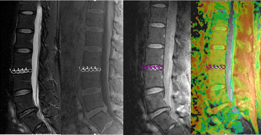
Fig.1MR images of the lumbar spine .Every IVD was cut into 5 uniform parts in each UTE,R2*and T1rho.
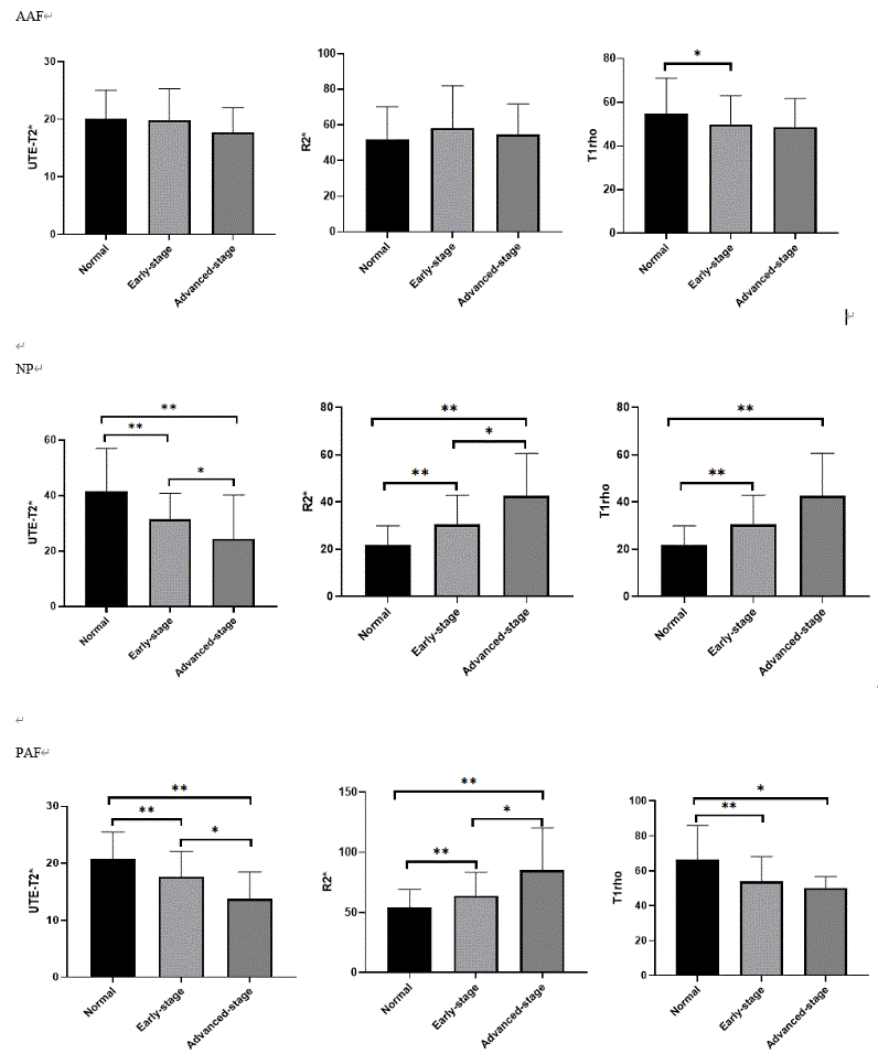
Fig.2 Post hoc multiple comparisons among three Disc degeneration groups
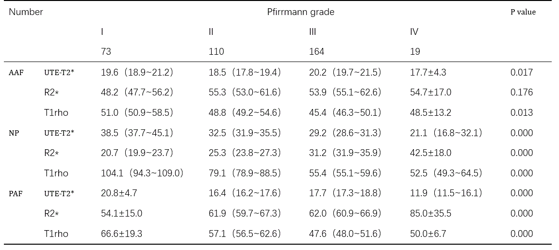
Table1 UTE-T2*、R2* and T1rho values of AAF, NP, and PAF with Pfirrmann grades