3158
Alterations in regional and network-level neural function in patients with HCV infection and its association with cognitive dysfunction1Department of Radiology, Fujian Medical University Union Hospital, Fuzhou, China, 2SIEMENS Healthcare, Shanghai, China
Synopsis
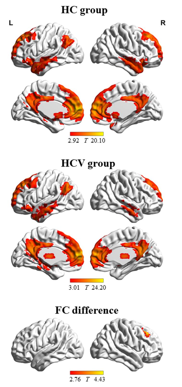
We aimed to investigate alterations in regional and network-level neural function in patients with hepatitis C virus (HCV) infection and examine the association between these alterations and patients’ cognition dysfunction. HCV-infected patients performed significantly worse in the cognitive tests. In the HCV group, amplitude of low-frequency fluctuation (ALFF) decreased in two prefrontal regions (Region-1and Region-2). The HCV group showed lower FC between the Region-1 and right middle frontal gyrus; whereas they presented an increase in FC between Region-2 and bilateral parietal-temporal cortex. HCV infection affects brain function, including local intrinsic neural activity and global functional integration.
Introduction
There is increasing evidence of the negative impacts of HCV infection on cognitive functions such as attention and executive deficits, memory problems, and learning disability.1 HCV infections can also induce alterations in spontaneous brain activity, including the slow down of the average dominant frequency of electroencephalogram (EEG) 2and the decrease of glucose metabolism in a few brain regions.3 Thus, resting state functional magnetic resonance imaging (fMRI) may be potentially utilized in the assessment of brain functional alterations that are related to HCV infection. Analysis of the amplitude of low-frequency fluctuation (ALFF) and seed-based functional connectivity (FC) was performed to respectively assess regional neural function and functional integration. ALFF reflects regional spontaneous cerebral neural activity,4 whereas FC reveals the functional coordination between various regions.5 This study aimed to investigate alterations in regional and network-level neural function in patients with HCV infection and examine the association between these alterations and patients’ cognition dysfunction.Methods
The study included 17 patients with HCV infection and 17 healthy controls. These individuals had undergone resting state fMRI as well as cognitive assessment using a battery of tests that was collectively called the “Psychometric Hepatic Encephalopathy Score (PHES)” examination. Analysis of ALFF and seed-based FC were conducted to respectively assess regional neural function and functional integration.Results
HCV-infected patients performed significantly worse in the cognitive tests. In the HCV group, ALFF decreased in Region-1 (left medial frontal gyrus and bilateral anterior cingulate gyrus) and Region-2 (right middle and superior frontal gyrus). The HCV group showed lower FC between the Region-1 and right middle frontal gyrus; whereas they presented an increase in FC between Region-2 and the left supramarginal gyrus/superior temporal gyrus and right supramarginal gyrus. No significant correlation was observed between ALFF/FC measurements and PHES result.Conclusion
This preliminary study presents additional evidence that HCV infection affects brain function, including local intrinsic neural activity and global functional integration.Acknowledgements
The National Natural Science Foundation of China (No. 81501450 and 82071900), Fujian Provincial Science Fund for Distinguished Young Scholars (No.2018J06023), Fujian Provincial Program for Distinguished Young Scholars (No. 2017B023), Fujian Provincial Program for Science and Technology Innovation (No.2019Y9067), Fujian Provincial Health Commission Project for Scientific Research Talents (No. 2018-ZQN-28), and Academic Exchange Fund for Postgraduates of Fujian Medical University supported this study.References
1. Thein HH, et al (2007) Improved cognitive function as a consequence of hepatitis C virus treatment. HIV Med 8:520-528.
2. Weissenborn K, et al (2004) Hepatitis C virus infection affects the brain-evidence from psychometric studies and magnetic resonance spectroscopy. Journal Of Hepatology 41:845-851.
3. Heeren M,et al (2011) Cerebral glucose utilisation in hepatitis C virus infection-associated encephalopathy. J Cereb Blood Flow Metab 31:2199-2208.
4. Wang Z, et al (2011) Spatial patterns of intrinsic brain activity in mild cognitive impairment and Alzheimer's disease: a resting-state functional MRI study. Human Brain Mapping 32:1720-1740.
5. Barkhof F, et al (2014) Resting-state functional MR imaging: a new window to the brain. Radiology 272:29-49.
Figures