3157
Altered cortical-striatal network in patients with hemifacial spasm1China-Japan Friendship Hospital, Beijing, China, 2GE healthcare, China, Beijing, China
Synopsis
Hemifacial spasm (HFS) is a motor disorder. The purpose of this study was to explore the alterations of the cortical-striatal network in HFS using resting-state fMRI. We found that the functional connectivity (FC) between the putamen and ventral striatum and both motor and emotion-related cortex was increased in patients with HFS compared to the healthy controls. The FC between the ventral striatum and the motor cortex was positively correlated with the spasm intensity. To sum up, HFS may lead to altered neural activity in the cortical-striatal loop.
Introduction
Hemifacial spasm (HFS) is a syndrome characterized by unilateral, involuntary contractions of the facial muscles innervated by the affected facial nerve1. Studies have found that depression and anxiety are more common in patients with HFS2, 3. Striatum plays a prominent role in modulating motor activity and higher cognitive function4. However, the exact neural mechanism of the striatum in the regulation of motor in HFS patients remain unexplored. Early identification of functional changes in the cortical-striatal loop of HFS patients can help understand disease pathogenesis and achieve early diagnosis and effective treatments. This study aimed to investigate if an altered cortical-striatal network exists in patients with primary unilateral HFS using resting-state fMRI.Methods
The fMRI data of 30 adult patients with primary unilateral HFS (left 15, right 15) and 30 healthy controls were collected using a 3.0T MRI scanner. The data of left-side HFS and matched controls were flipped from left to right, and then the right side was considered as the ipsilateral side. Six subregions of the striatum in each hemisphere were selected as seeds for FC analysis. The intra-group FC of the cortical-striatal network was analyzed by a one-sample t-test. The two-sample t-test was performed to compare the difference of FC in the cortical-striatal network between the two groups (GRF correction, voxel P < 0.005, cluster P < 0.05). The psychological status of all subjects was assessed by the self-rating depression scale (SDS), and the spasm intensity of HFS patients was evaluated by the Cohen spasm score. Then, the correlations between FC and both Cohen spasm score and SDS score were analyzed.Results
The intra-group FC maps of the cortical-striatal network in the healthy and patient groups were similar, and the between-group FC of the cortical-striatal network in the two groups showed significant differences. In HFS patients, the FC between dorsal rostral putamen and ventral striatum and both motor and emotion cortex was increased (corrected P < 0.05), while between the dorsal caudal putamen and inferior ventral striatum and ipsilateral motor cortex was decreased (corrected P < 0.05) (Fig. 1). Furthermore, the FC between the ventral striatum and motor cortex was positively correlated with spasm intensity. No correlation was found in the controls.Conclusions
Primary unilateral HFS induces several FC alterations in the cortical-striatal network. The spasm intensity of facial muscles is associated with these functional alterations. This study indicated HFS caused increased neural activities between the striatal subregions and motor and emotion cortex. Early detection of functional changes broadens the understanding of the disease pathogenesis and consequently boosts the possibility of early diagnosis and treatment.Acknowledgements
This study was supported by the National Natural Science Foundation of China (NSFC) (Nos. 81971585), and The National Key Research and Development Program of China (Nos. 2020YFC2003903).References
1. Palacios E, Breaux J, Alvernia J E. Hemifacial spasm. Ear Nose Throat J. 2008, 87: 368.
2. Huang Y-C, Fan J-Y, Ro L-S, et al. Validation of a Chinese version of disease specific quality of life scale (HFS-36) for hemifacial spasm in Taiwan. Health Qual. Life Outcomes. 2009, 7: 104.
3. Rudzinska M, Wojcik M, Malec M, et al. Factors affecting the quality of life in hemifacial spasm patients. Neurol. Neurochir. Pol. 2012, 46: 121-9.
4. Rosen G D, Williams R W. Complex trait analysis of the mouse striatum: independent QTLs modulate volume and neuron number. BMC Neurosci. 2001, 2: 5.
Figures
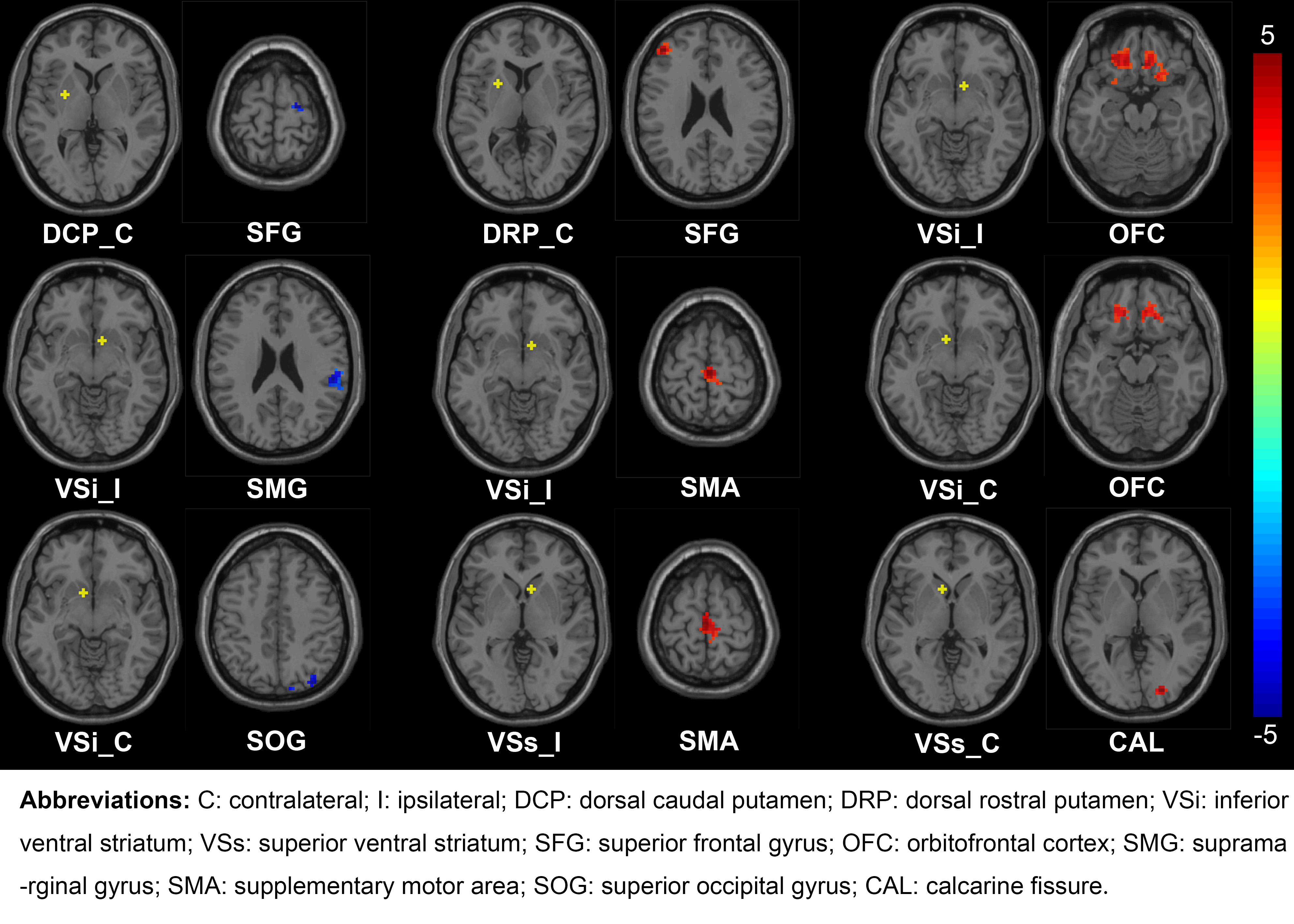
Fig. 1: Differences of the cortical-striatal FC in patients with HFS compared to healthy controls. The FC between striatal subregions and both motor and emotion-related cortex was statistically different between the two groups (GRF correction, voxel P < 0.005, cluster P < 0.05). The yellow dots in the brain represent the seeds of striatal subregions. The red regions represent the increased FC between seeds and cortexes, while the blue areas represent a decreased FC. The color bar represents the t value.