3148
Tracing effects of breath-training on healthy brains using RS-fMRI1National Magnetic Resonance Research Center (UMRAM), Bilkent University, Ankara, Turkey, 2The Faculty of Physical Therapy and Rehabilitation, Hacettepe University, Ankara, Turkey, 3Faculty of Medicine, Department of Radiology, Hacettepe University, Ankara, Turkey
Synopsis
The present study was conducted to investigate the effects of Inspiratory Muscle Training (IMT) on brain activity in healthy people using resting state functional magnetic resonance imaging (RS-fMRI). Participants were assigned into a training group (n=14), which consisted of 2 subgroups, namely sham group (n=7) and treatment group (n=7) and their native a baseline (n=14) group. Apart from global correlations, brain intrinsic networks differences between groups and subgroups were also examined. Compared to the baseline and sham there was a decrease in global BOLD signal changes and more localized clusters of activation in the treatment group.
Purpose
Inspiratory muscle training (IMT) plays a role in increasing respiratory muscle strength and we aimed to investigate the effects of IMT on brain activity in healthy people.Methods
This was a prospective, randomized controlled study. IRB approval was obtained for this study and all participants signed a consent form.Subjects
Fourteen right handed healthy subjects (8F/6M, mean age:28.14 ±5.246) without any history of systemic, neurolo psychiatric disorders or any kind of drug with possible effects on CNS or cardiopulmonary system constituted ‘training’ group. RS-fMRI was performed for all 14 participants, then they were randomly assigned to sham group (n=7, 4F/3M, 29.57 ±7.208 years) and treatment group (n=7, 4F/3M, mean:26.71±1.704 years). Following 8 weeks breath training, both groups underwent RS-fMRI again.
Breath Training
The maximal inspiratory pressure (MIP) and maximal expiratory pressure (MEP) were assessed using a mouth pressure device (MicroRPM, Micromedical, Kent, UK). The MIP was measured near a residual volume after a maximal expiration, and the MEP was measured near total lung capacity after a maximal inspiration. Tests were repeated until no further improvements were obtained and there was less than a 10% difference between the two best values. Participants were trained using an inspiratory threshold-loading device (POWERbreathe, Southam, UK). The treatment group received IMT at 60% of MIP. The MIP was measured every week, and the resistance was adjusted to maintain 60% of MIP. The sham group received sham IMT at a fixed workload of 15% of MIP. Both groups trained for 30 minutes per day, 5 days per week, for 8 weeks.
Image Acquisition
RS-fMRI was performed with a 3.0-T scanner (Siemens Trio, Germany) magnet equipped with 32 channel phase array head coil. A Gre EPI sequence ( TR/TE,2000/34 ms; 184 volumes) was obtained while the subjects were lying quietly inside the scanner. A 3D T1W-MPRAGE (TR/TE,2600/3.02 ms; 176 volumes with 1 mm slice thickness) was also obtained from each subject.
Data Processing and Analysis
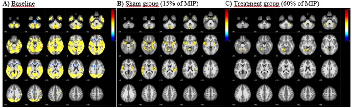
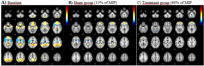
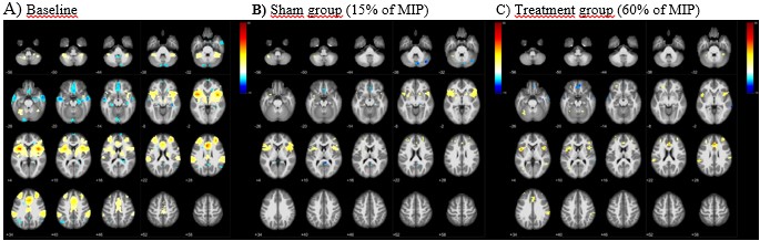
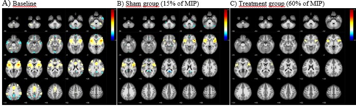
RS-fMRI data preprocessing and analyses were obtained with CONN toolbox1 implemented in Matlab20202. For the reduction of noise in BOLD data Component Based Noise Correction method (CompCor) was applied. For MNI coordinates and anatomical labeling SPM123 and bspmview toolbox4 were used. Global correlations were performed as the average of correlation coefficients between each individual voxel and all of the voxels in the brain (voxel threshod p<.001 and cluster threshold p<.05). For brain intrinsic networks, seed based connectivity (SBC) was applied to the data which characterizes the connectivity patterns with a pre-defined seed. These networks are DMN (medial prefrontal cortex (MPFC), left and right lateral parietal (LP) and posterior cingulate cortex (PCC), salience network (Anterior Cingulate Cortex (ACC), Insula (L-R), rostral prefrontal cortex (L-R) (RPFC), supramarginal gyrus (L-R) (SMG)) and Insula network (L and R Insula). The color scale represents the t value of the BOLD signal.
Results
Both groups were similar in terms of age, gender and education (p>.05). The results show the whole-brain average global BOLD and connectivity alterations in three important networks (Default Mode Network (DMN), Salience and Insula Network) in the brain related to such as self-referential processing-interoception, communication-social-behavior-self-awareness, consciousness-homeostasis respectively. There are also changes in both activation clusters and activation strength when compared with baseline.ACC activation disappeared in sham group compared to baseline while activation is seen in ACC 5 for the treatment group compared to the sham (Fig.3).
Conclusion
After 8 weeks breath training, the treatment IMT shows less activation clusters with less activation strength compared to the sham IMT. Our results agree with previous meditation studies including breathing training, showing that DMN is deactivated and network gets more organised and localised6,7. Breath training at 60% IMT was more affective on respiratory related areas, attention processing and motor control areas of the brain than sham IMT which points to increased respiratory awareness in healthy people.Acknowledgements
No acknowledgement found.References
1. Conn: A functional connectivity toolbox for correlated and anticorrelated brain networks. Brain connectivity, 2(3), 125-141)
2. Mathworks Inc., Natick MA, USA
3. https://www.fil.ion.ucl.ac.uk/spm/software/spm12/
4. https://www.bobspunt.com/bspmview/
5. Hölzel B K., Ott U, Hempel H, Hackl A, et al. D. 2007. Differential engagement of anterior cingulate and adjacent medial frontal cortex in adept meditators and non-meditators.
6. Brewera J A, Worhunskya P D, Grayb J R, et al. 2011. Meditation experience is associated with differences in default mode network activity and connectivity.
7. Doll A, Hölzel B K, Boucard C C, et al. 2015. Mindfulness is associated with intrinsic functional connectivity between default mode and salience networks.
Figures