3147
The effects of one rTMS session on the left DLPFC on episodic future thinking: preliminary results.1Radiology, UZ Brussel (VUB), Brussels, Belgium, 2Head and Skin, UGent, Ghent, Belgium, 3Psychiatry, UZ Brussel (VUB), Brussels, Belgium, 4Laboratoire de Neurosciences Intégratives et Cliniques, Université de Bourgogne Franche-Comté, Bourgogne, France
Synopsis
Episodic future thinking (EFT) is the ability to imagine events to occur in one’s personal future. In this study, we studied the interaction of one rTMS session on the left DLPFC with EFT on the connectivity between areas from the DMN, the hippocampus and the amygdalae.
All participants performed an EFT task before and 1 hour after a real or sham rTMS session during a fMRI scan.
Our preliminary results showed interesting changes in the connectivity between the MPFC and right LP with both amygdalae. However, more data is needed to strengthen these results.
Introduction
Episodic future thinking (EFT) is the ability to imagine events to occur in one’s personal future1. EFT processing is mainly done by areas from the default mode network (DMN), the dorsolateral prefrontal cortex (DLPFC) and hippocampus1.To gain more insight in the treatment effects of repetitive transcranial magnetic stimulation (rTMS) on the left DLPFC in depression, the effect of one rTMS session on cognitive functions is studied in healthy volunteers2. However, the existence of any effect of 1 rTMS session in healthy subjects is still debated2.
In the current study, the effect of one rTMS session in healthy subjects on EFT was studied with fMRI.
Methods
23 healthy subjects (female:17, age:24±3) participated in the study. All scans were done on a 3T GE MRI scanner. For each individual, the rTMS coil position to target the left DLPFC was determined based on a 3D T1 weighted head scan3 and the stimulation intensity was determined based on the threshold to evoke a motoric response (MT). 12 subjects received real rTMS stimulation at 110% MT and 11 subjects received sham stimulation at 10% MT. The rTMS was given while the participants remained inside the MRI scanner using a Magstim rapid stimulator and a MRI compatible coil. The stimulation was done in 3 blocks with 5 minutes in between. Each block contained 10 trains of 2s stimulation at 20Hz and 12s rest.Before and 1 hour after the rTMS session, the subjects performed an EFT task during a fMRI scan (T/R head coil, TR/TE=2/0.30s, 3.75x3.75x4mm resolution, 207 dynamics). The task consisted of 18 trials in which a word was presented for 20s while the participant was thinking of an associated event possibly to occur in its own future. We instructed the events to be concrete, realistic and limited in time and space. Once an event was found, the participants had to push a button.
Since, we used a fMRI task with only 1 continuous state, we processed the data following a resting state pipeline in SPM124 and CONN5. We compared the connectivity between areas of the DMN, the hippocampus and the amygdalae before and after a real or sham stimulation using a ROI-to-ROI analysis.
Results
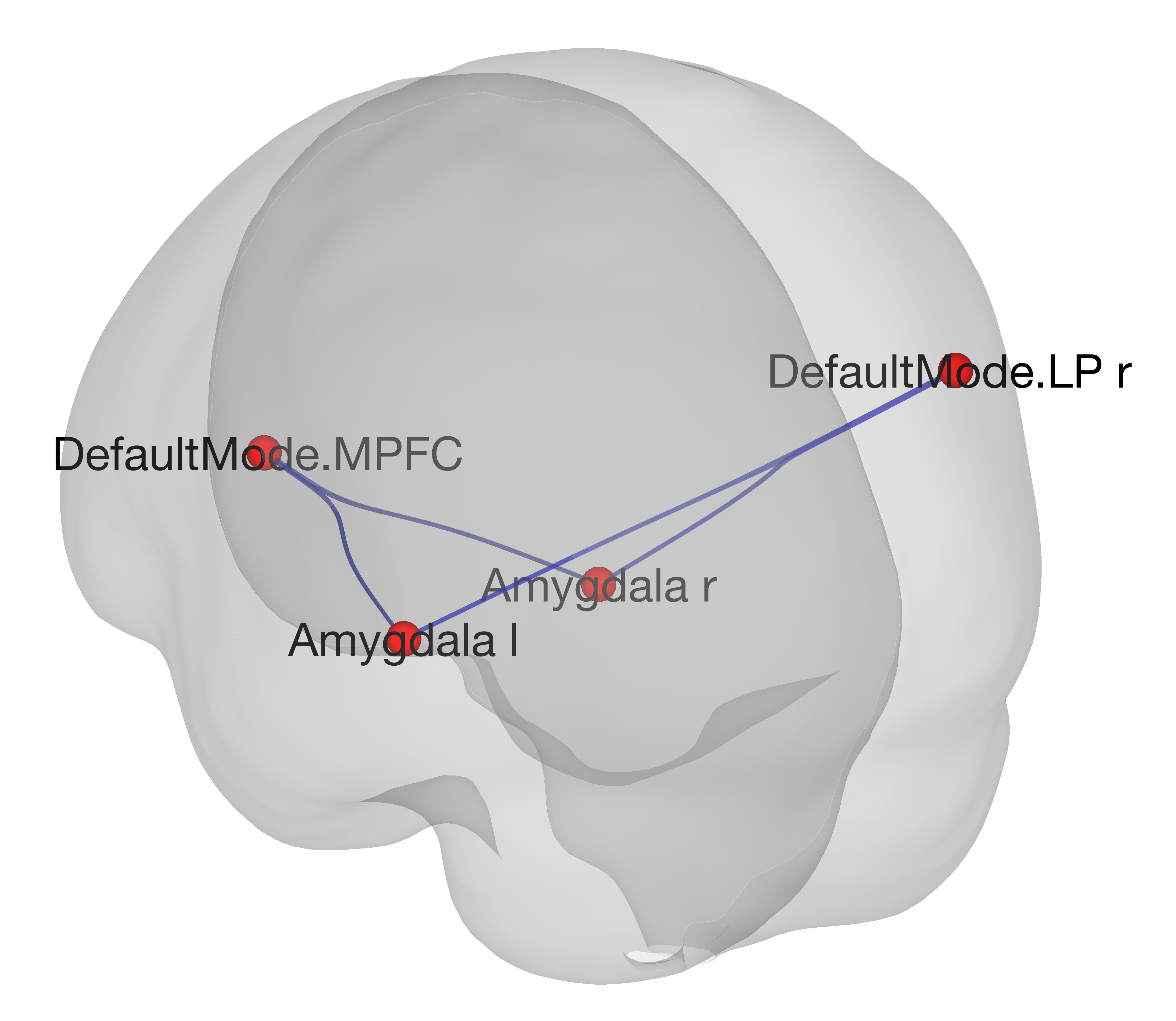
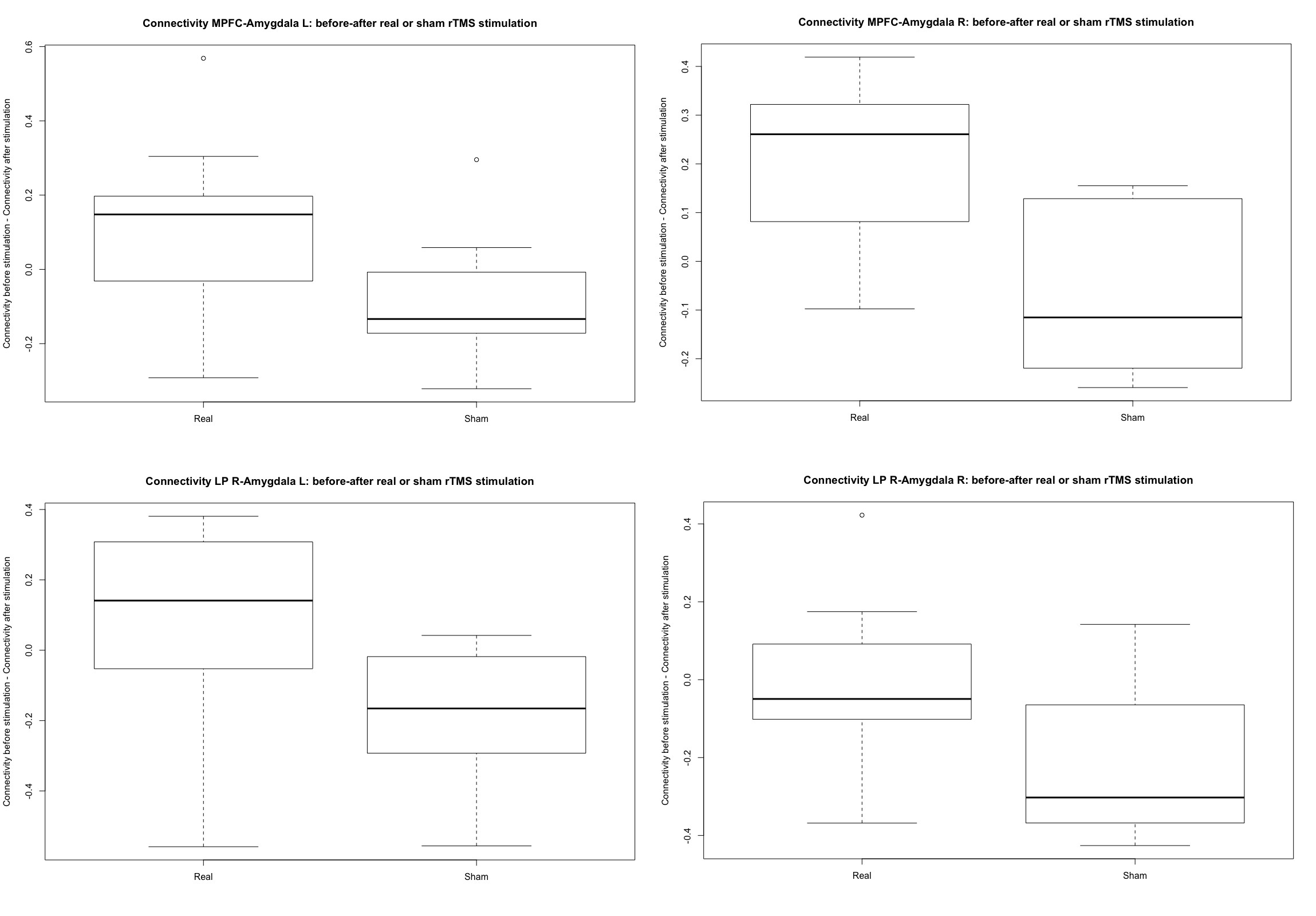
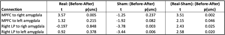
No significant effect was found of stimulation type (real or sham) on the difference in response time to find a future event, between the before and after session (F=0.53, p=0.475).Within the selected ROIs, a significant interaction (Real – Sham stimulation) x (Before – After stimulation) was found in a network formed by the left and right amygdala, the medial prefrontal cortex (MPFC) and the right lateral parietal cortex (LP) (Fig. 1) (Mass=59.99, pFWE=0.043 (FWE corrected at network level)). Post-hoc tests per connection, revealed a decreased connectivity between the MPFC and the right amygdala after real stimulation and an increased connectivity between the right LP and both amygdalae after sham stimulation (table 1 and fig. 2).
Discussion
Although, the sample size in this intermediate analysis of our rTMS-fMRI combined study is still small, interesting changes in the connectivity between areas of the DMN and the amygdalae during EFT were found. The most significant effect of rTMS stimulation observed, was a decreased connectivity between the MPFC and the right amygdala. This finding is of interest since it is shown that through the coupling of the MPFC with the hippocampus and amygdala, EFT affect valuing the reward of possible future events, and hence, the decisions we make regarding personal preferences and goals6,7. However, our EFT task did not involve a future reward evaluation.In the review study of Patel et al.2, based on behavioral studies, 1 excitatory (>1Hz) rTMS session on the left DLPFC was found to have no effect on the results of an episodic memory task performed before and after the stimulation. Episodic memory and EFT are closely related cognitive functions sharing the same neural network1. In general, the lack of any effect on the behavioral results of cognitive studies was opposite to reported changes in the connectivity in meso-cortico-limbic networks1.
After acquiring more data, it would be of interest to perform a more detailed analysis of the dynamic and causal connectivity between the MPFC and the amygdala during EFT and the modulating effect of rTMS delivered on the left DLPFC, on these interactions.
Conclusion
A preliminary analysis of our data revealed interesting effects of 1 rTMS session on the left DLPFC, on the connectivity between the MPFC and right LP with both amygdalae. However, more data is needed to confirm and strengthen these results.Acknowledgements
No acknowledgement found.References
1. Schacter D.L., Benoit R.G., Szpunar K.K. Episodic Future Thinking: Mechanisms and Functions. Curr. Opin. Behav. Sci. 2018;17:41-50
2. Patel R., Silla F., Pierce S. et al. Cognitive functioning before and after repetitive transcranial magnetic stimulation (rTMS): A quantitative meta-analysis in healthy adults. Neuropsychologia 2020;141:107395
3. Peleman K., Van Schuerbeek P., Luypaert R. et al. Using 3D-MRI to localize the dorsolateral prefrontal cortex in TMS research. World J. Biol. Psychiatry 2010;11( 2 P 2):425-430
4. https://www.fil.ion.ucl.ac.uk/spm/
5. www.nitrc.org/projects/conn, RRID:SCR_009550
6. Stawarczyk D., D’Argembeau A. Neural correlates of personal goal processing during episodic future thinking and mind‐wandering: An ALE meta‐analysis. Hum. Brain Mapp. 2015;36(8):2928-2947
7. Peters J., Buchel C. Episodic future thinking reduces reward delay discounting through an enhancement of prefrontal-mediotemporal interactions. Neuron 2010;66(1):138-148
Figures