3120
Initial Experience of Body Imaging at 5T
Zhenhua Shen1, Xuchen Zhu1, Shihong Han1, Fuyi Fang1, Wei Luo1, Shao Che1, Zidong Wei1, Jinguang Zong1, Yongquan Ye2, Bo Li1, Shuheng Zhang1, Anthony Vu2, Weiguo Zhang2, and Guobin Li1
1United Imaging Healthcare, Shanghai, China, 2UIH America, Inc., Houston, TX, United States
1United Imaging Healthcare, Shanghai, China, 2UIH America, Inc., Houston, TX, United States
Synopsis
For the first time, routine clinical body imaging with large FOV on a whole-body 5T MRI system is demonstrated. With multi-channel RF parallel transmission hardware architecture and static RF shimming techniques, the uniformity of the RF transmission field is shown to be well controlled for imaging quality guarantee. Preliminary results show great promise for body imaging at 5T.
Introduction
Whole body MR imaging at ultra-high field remains challenging due to transmit B1(B1+) field inhomogeneity, as radiofrequency (RF) wavelength in human tissues is much shorter than the dimensions of human torso1 and causing constructive/destructive interference patterns in resultant images. In order to mitigate the issue, RF shimming methods including static parallel transmission (static pTx, or static RF shimming) and dynamic parallel transmission (dynamic pTx, or dynamic RF shimming) have been proposed on the basis of multi-channel transmission hardware architecture.2 Here we present the initial results of abdomen and pelvis imaging at 5T with static RF shimming, and demonstrate the capability of the 5T MR system for routine body imaging.Methods
Experiments were performed on a whole body 5T MRI scanner (United Imaging Healthcare, Shanghai, China) as shown in Figure 1. The parallel RF transmission system consists of eight independent RF power amplifiers (RFPAs), each with a peak power of 8kW. The volume transmission coil used was an eight-channel loop array coil. For the initial testing, only the amplitude and phase of each channel were changed to optimize the target B1+ fields which is commonly used in 3T system as static RF shimming method. The static RF shimming procedure starts with a prescan calibration step with a B1+ mapping sequence. The total acquisition time for all eight channels in prescan takes less than 1 minute. Desired weights of each transmit channel are then calculated using magnitude least squares optimization and used for the later scan.3 The T2-weighted (T2W) fast spin echo (FSE) sequence with fat saturation (FOV: 380mm*300mm, Matrix: 336*225, TR: 4000ms, TE: 87.2ms, Slice Thickness: 6mm, Slice Number: 24) was selected as the demonstrating protocol for abdominal imaging (Figure 2). Another routine T2W FSE sequence (FOV: 350mm*350mm, Matrix: 416*312, TR: 4800ms, TE: 75.04ms, Slice Thickness: 3mm, Slice Number: 26) was chosen for image quality assessment of pelvic imaging (Figure 3).Results
Figure 2 and Figure 3 demonstrate the B1+ profiles of both circular polarization (CP) mode and optimized transmitting mode used after static RF shimming process in abdomen and pelvis. The coefficient of variance (CV) reflects the degree of B1 homogeneity, and greater CV values correspond to greater inhomogeneity. Before static RF shimming, severe shading artifacts can be seen in T2W FSE images. After static RF shimming, B1+ uniformity within the abdomen and pelvis was improved to yield smaller CV values, and the resultant image became more uniform. The CV value decreased by nearly 16% compared with that of CP mode in abdomen, and by 33% in pelvis.Conclusion
Preliminary results show great promise for routine whole body MR clinical imaging at 5T, an area where 7T has been struggling due to more severe B1+ inhomogeneity and SAR constraints. The static RF shimming procedure takes less than a minute and can be implemented as part of the prescan procedures. Further improvements, such as by dynamic RF shimming, will be investigated in future study.Acknowledgements
This work is supported by National Key R&D Program of China N0.2017YFC0108800.References
- Erturk, M. A.; Li, X.; Van de Moortele, P. F.; Ugurbil, K.; Metzger, G. J., Evolution of UHF Body Imaging in the Human Torso at 7T: Technology, Applications, and Future Directions. Top Magn Reson Imaging 2019, 28 (3), 101-124.
- Padormo, F.; Beqiri, A.; Hajnal, J. V.; Malik, S. J., Parallel
transmission for ultrahigh-field imaging. NMR
Biomed 2016, 29 (9), 1145-61.
- Setsompop, K.; Wald, L. L.; Alagappan, V.; Gagoski, B. A.; Adalsteinsson, E., Magnitude least squares optimization for parallel radio frequency excitation design demonstrated at 7 Tesla with eight channels. Magn Reson Med 2008, 59 (4), 908-15.
Figures
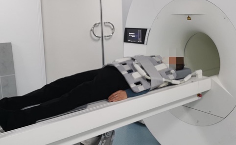
Figure 1 Photo of a healthy volunteer ready for abdominal scanning.
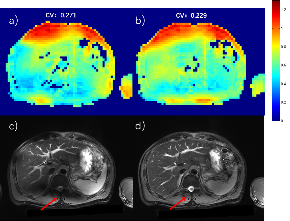
Figure 2 Abdominal imaging at 5T. a) and c) B1+ sensitivity map and T2W FSE image with CP mode excitation. b) and d) B1+sensitivity map and T2W FSE image after static RF shimming optimization. The CV values were measured for bothcases.The red arrows show the most significant improvement area.
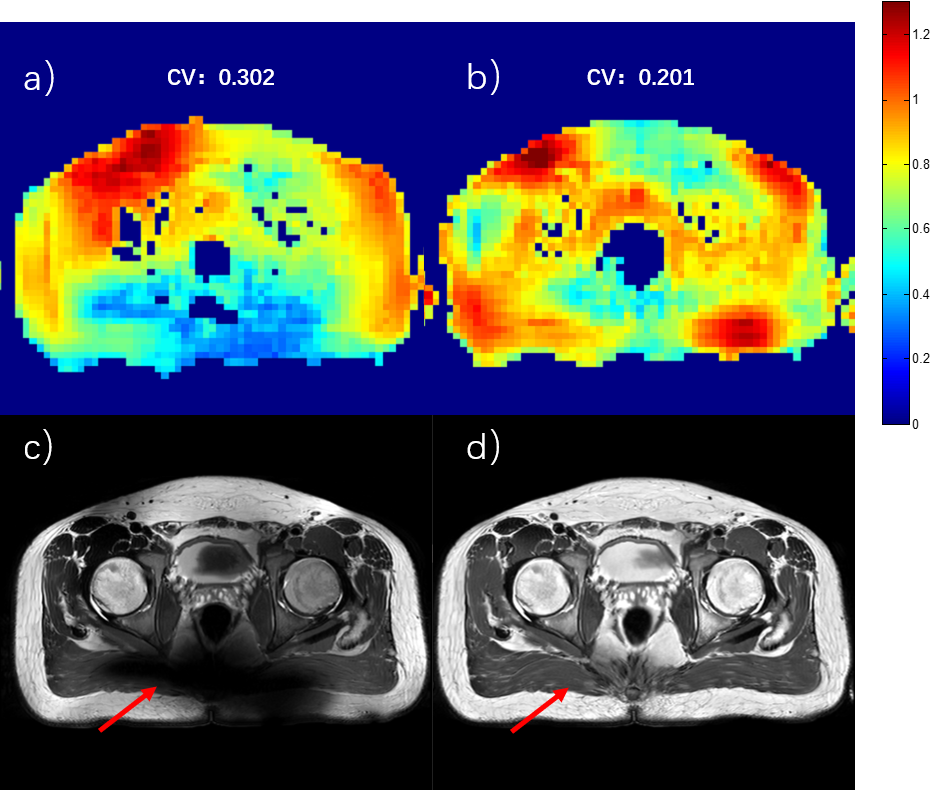
Figure 3 Pelvic imaging at 5T. a) and c) B1+ sensitivity map and T2W FSE image with CP mode excitation. b) and d) B1+sensitivity map and T2W FSE image after static RF shimming optimization. The CV values were measured for bothcases.The red arrows show the most significant improvement area.