3118
Very low field MRI for brain imaging1SPEC - CEA Saclay - Université Paris Saclay, Saclay, France
Synopsis
Classic high field MRI is a powerful tool in healthcare. However, it is an expensive and complex installation and it might be useful to supply a less convenient device adapted to a lot of usage not requiring high field potential. We have developed a very low field MRI working at between 1mT and 10mT. Main static field is providing by a water-cooled resistive magnet. In order to achieve an iso spatial resolution of 2mm in a reasonably short time of acquisition, we have worked on adapted sequences including compressed sensing and optimized detection with parallel acquisition.
Introduction
Very low field MRI (VLFMRI) present several advantages compared to high fields: it is very cheap, easier to install, silent and can accept metallic objects which allows to accept population presently excluded to high field MRI. However, the main drawback is the weakness of the signal (150 times lower than at 1.5T) and the usually poor sensitivity of tuned coils at low frequencies.With ideal magnetic sensors, this factor 150 is partly compensated by to the reduction of the acquisition bandwidth, the short T1 at low fields and the lower body noise. Then the cost in signal to noise at 10mT is between 4 to 8 compared to 1.5T which is less than 2 in iso resolution.
Hence, VLFMRI requires first a very good external shielding and an optimized detection. In addition, we have also to adapt sequences used at high field to low field to improve the exam duration.
Materials and methods
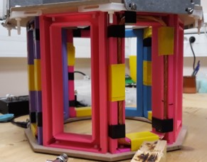
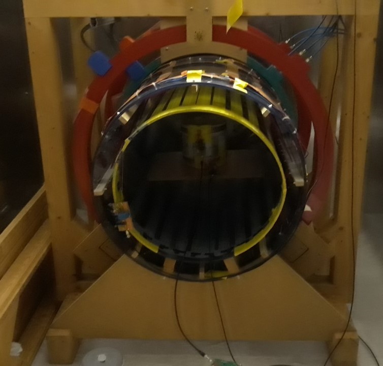
Our system1 is composed of resistive coils with two different configurations. One based on Gareth model with 6 coils is very homogeneous but is rather closed and dedicated to lying people. A second configuration with 3 coils rather opened can accept sitting people but is less homogeneous, It reaches about 100ppm on a field of view of 20cm3. Radiofrequency pulses are generated by a 5 elements large saddle shaped coil, differentiated and decoupled from sensor. We have developed a parallel acquisition (4 to 8 channels) with two types of detection. A room temperature tuned coils optimized to this field and mixed superconducting-magnetoresistive sensors at present slightly less performing but offering a wide frequency detection useful for field sweeping.At room temperature, channel sensor are windings litz coil (figure 1). The channel sensibility is 0.2 fT/√Hz and quality factor Q is about 150. The quality of the tuning capacitance is critical to achieve high quality factors.
For the sequences, we have explored different approaches to optimize the acquisition duration. It is important to note that classical EPI or radial sequences with slice selection cannot by efficiently applied at low fields as slice selection would require either long soft pulses, either very high gradients not compatible with very low fields. Double encoding is hence required, inducing a rather long computation time for non Cartesian k space covering.
In terms of k-space covering it is also important to note that a large number of multi-echos cannot be used as acquisition time which is related to the resolution targeted is rather long (20-30ms). So exam acceleration requires to reduce the number of trajectories in k-space. We are presently investigating the use of Sparkling techniques to reduce this number of trajectories while maintaining a good spatial resolution of the images.
Channels sensor are 8 windings litz coil. The channel sensibility is 0.2 fT/√Hz and quality factor Q is about 150, which is as closed as we are allowed to reach, because of the working bandwidth (about 4 kHz).
Results
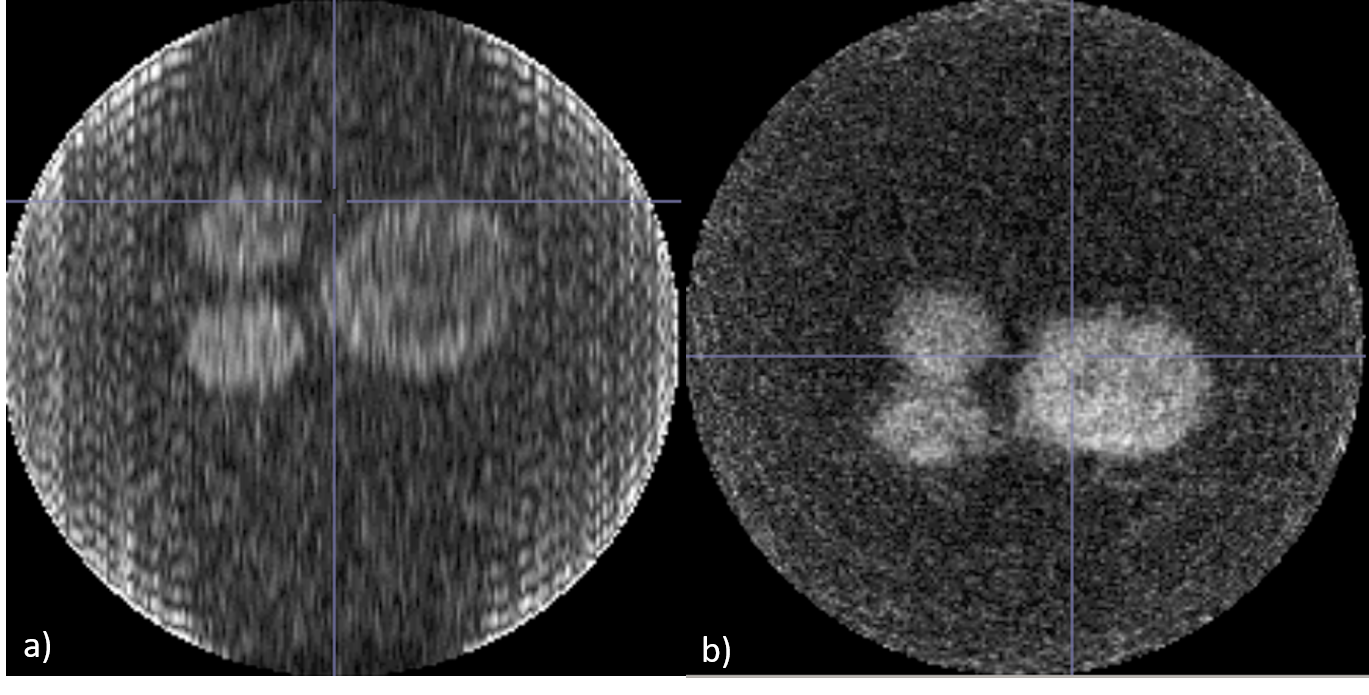
We are now able to process 3D images on phantom (three bottles filled with NiCl doped water) with cartesian (as reference) and radial sequence (Figure 3). It takes about half an hour to get the first with a resolution of 2*2*0.75mm, and a dozen of minutes to get the last one with a resolution of 2*0,75*0,75mm. It is important to notice that it is a radial stack of stars sequence, in the end we expect to shorten acquisition time with full 3D radial sequence.Conclusion
The signal to noise in our system is near theoretical one and allows us to acquire good quality images in a reasonable acquisition time. The next step is to implement efficient compressed sensing methods based on sparkling techniques2 to achieve clinical images in less than 4 minutes and in vivo tests.Acknowledgements
This work is supported by ANR found and CEA CFR scholarship.References
1. Herreros Q. Very Low Field Magnetic Resonance Imaging. :159.
2. Lazarus C, Weiss P, Vignaud A, Ciuciu P. An Empirical Study of the Maximum Degree of Undersampling in Compressed Sensing for T2*-weighted MRI. Magnetic Resonance Imaging. Published online 2018:1-31.
Figures