3110
Improving MRI Near Metal with Local B0 Shimming using a Unified Shim-RF Coil (UNIC): First Case Study, Hip Prosthesis in Phantom.1Biomedical Imaging Research Institute, Cedars-Sinai Medical Center, Los Angeles, CA, United States, 2Department of Bioengineering, UCLA, Los Angeles, CA, United States, 3Siemens Medical Solutions USA, Inc., Los Angeles, CA, United States, 4University of California, San Diego, San Diego, CA, United States, 5VA Medical Center, San Diego, CA, United States, 6University of Southern California Department of Radiology, Los Angeles, CA, United States
Synopsis
Metal artifact image degradation can interfere with tissue status assessment. Metal artifact reduction pulse sequences, mainly spin-echo based, and scanner B0-shimming can mitigate the artifacts. We have tested in a metal hip prosthesis phantom improving shimming locally with a novel technology that integrates separate but co-planar RF receive and shim arrays, expanding the freedom to position shim coils and shape B0, independently of the RF coil geometry and pulse sequence. Metal-induced signal-void artifact was reduced in GRE scans, revealing up to 50% additional area around the prosthesis. The sequence-agnostic technology may help improve MRI near metal prostheses and interventional devices.
Introduction
Clinical imaging near metal implants has advanced mainly through metal artifact reduction pulse sequences (MARS) such as SEMAC [1], MAVRIC [2], VAT [3], WARP [4], and MARS [5]. We propose to apply a novel shim-RF array, for the first time, to improve near-metal imaging which may have advantages: 1) Synergistically complementary to them, it may possibly improve all clinical MARS performance and scan times. 2) Compatible with any other sequence, e.g., GRE, UTE. 3) Unlike MARS, no tradeoffs in scan time, SNR, or SAR, especially important at 3T. Recently, localized multi-coil B0 shimming [6-10] has been demonstrated to go beyond the 2nd order shimming capability of scanners. We present a unified shim-RF coil (UNIC) design that can provide a stronger shim field to counteract the severe B0 inhomogeneity around metal, and is clinically viable in-part by integrating shimming with a conventional RF coil without SNR loss. Metal artifacts including signal loss can be especially severe in GRE sequences, avoided in imaging near metal implants [11] and depriving clinicians from GRE’s myriad advantages. For example, spin echo (SE) based clinical protocols recommended for imaging the hip or knee post-arthroplasty can last 30 to 50 minutes [12, 13]. On the other hand, SE-based sequences may sometimes not be an option, e.g., MR angiography near metal implants [12], or dynamic time-resolved studies. Improving B0 shimming such that some GRE sequences too may be reliably used near metal, can bring the benefits of GRE to bear in clinic. We report and quantify reduction in the signal void artifact and improvement in B0 homogeneity near the femoral head and stem of a metal hip prosthesis in phantom using a GRE sequence with local shimming.Materials and Methods
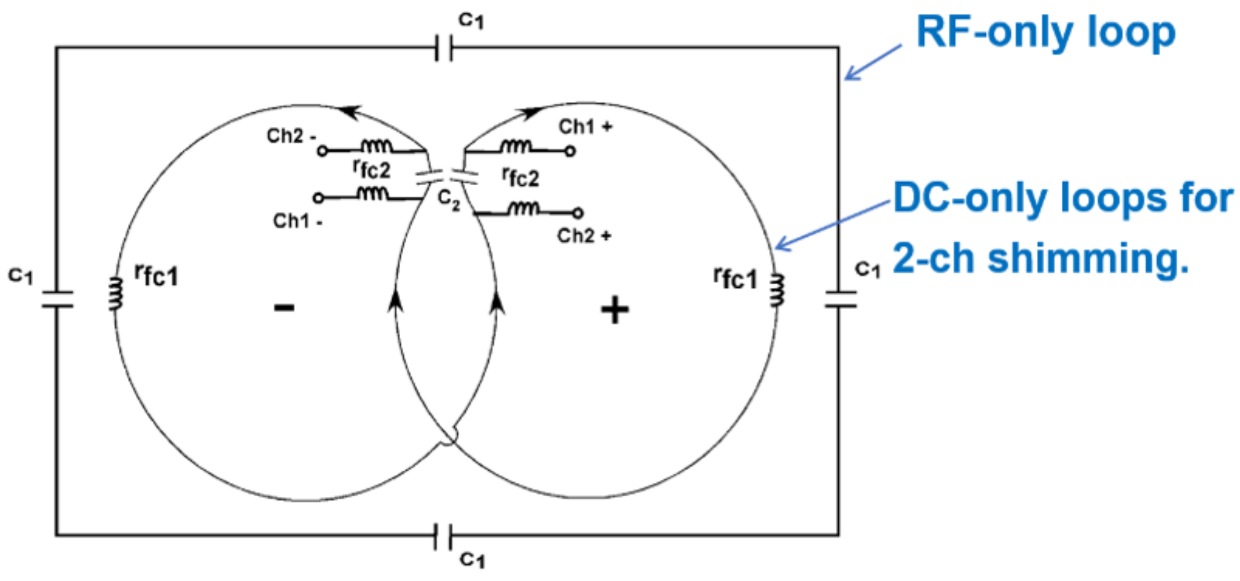
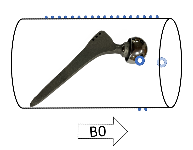
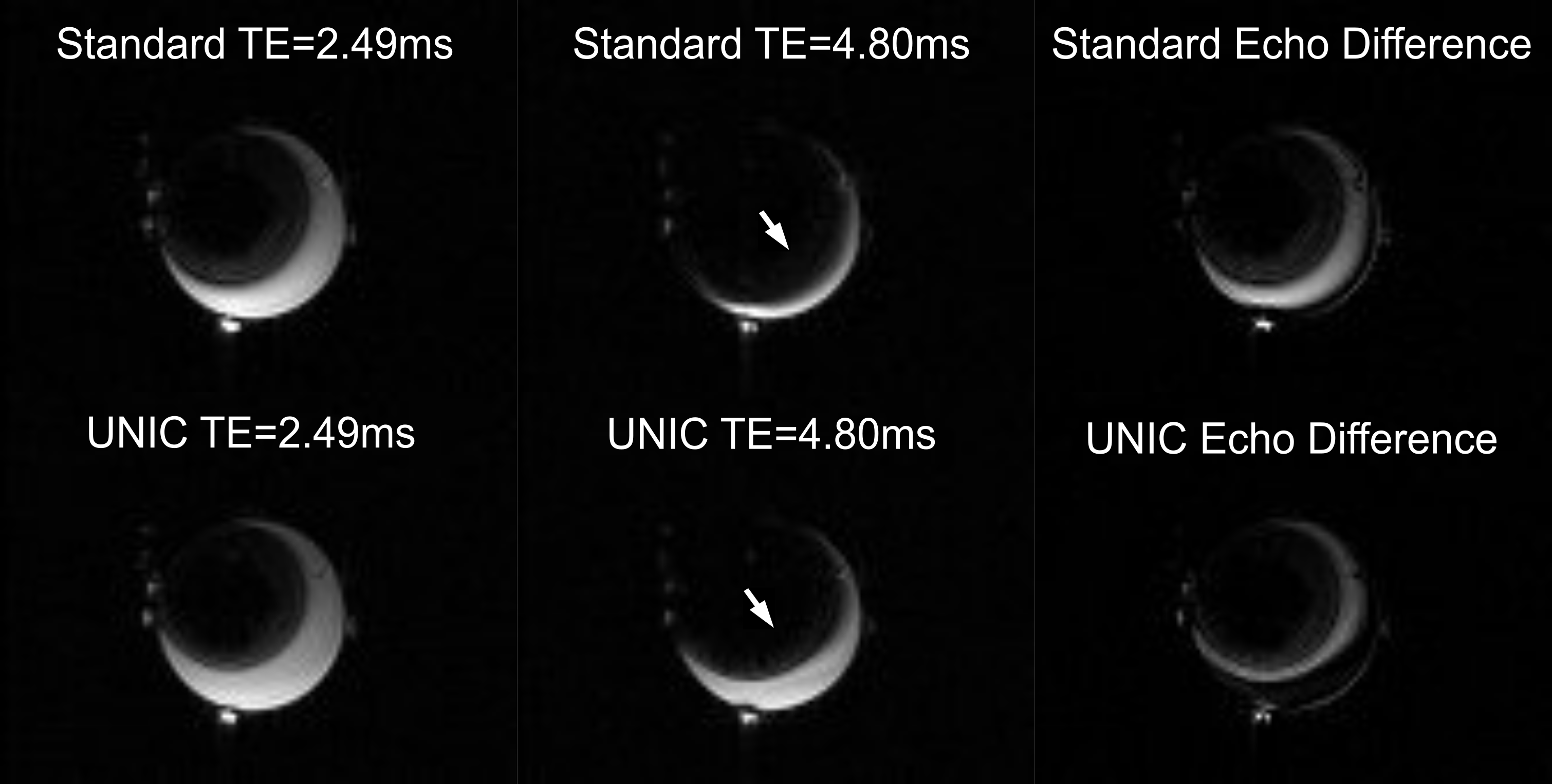
All scans were performed on a Siemens Biograph mMR 3-T scanner. A unified body coil [14-16] comprising two flat arrays (anterior and posterior) was constructed with 12 RF receive and 42 shimming channels with 2-turn loops in each shim coil, which doubled the shim field strength versus the prior [7]. The coil circuit design is depicted in Figure 1. Compared to the separate multi-coil shim approach [6], the unified shim-RF design can minimize the distance between shim coils and the image volume, thus greatly increasing shim efficiency and efficacy while also reducing power consumption and heat. The phantom, illustrated in Figure 2, was a near-cylindrical plastic container 23cm long and 12cm in diameter, containing a metal-on-metal cobalt-chrome hip prosthesis, femoral head 40mm diameter, femoral stem 155mm long, embedded in agarose gel. Fiducial markers were placed on the outside to help assess if UNIC shimming introduced signal displacement in the slice-encoding direction. First a baseline 3D double echo GRE scan was obtained with the scanner shim applied. Next, UNIC shim currents (limits +/-7A per channel) were computed in Matlab [17] using a least-squares fitting algorithm, and applied without the need for cooling, together with the scanner shim. The post-shim scan followed, with the same sequence/parameters as for the baseline: FOV = 300 mm x 225mm; image matrix = 112 x 84 x 60 (frequency x phase x partition); image resolution = 3.6 mm x 3.6 mm, axial partition (slice) thickness = 5.8 mm; TE1 = 2.49 ms , TE2 = 4.80 ms, TR = 517 ms, bandwidth = 795 Hz/pixel. Changes in signal void and visible areas around the metal were quantified using Matlab and ImageJ [18] by manually carefully drawing regions of interest (ROI) on 116 images (4 per slice, before and after UNIC shimming at both TE’s), and measuring the areas [2]. In images with the largest signal voids, ROI’s were also defined using ellipses or circles to reduce subjectivity due to phantom boundary ambiguity; then reported improvements are the more conservative (smaller) values.Results and Discussion
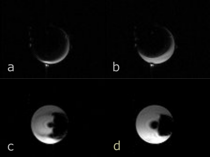
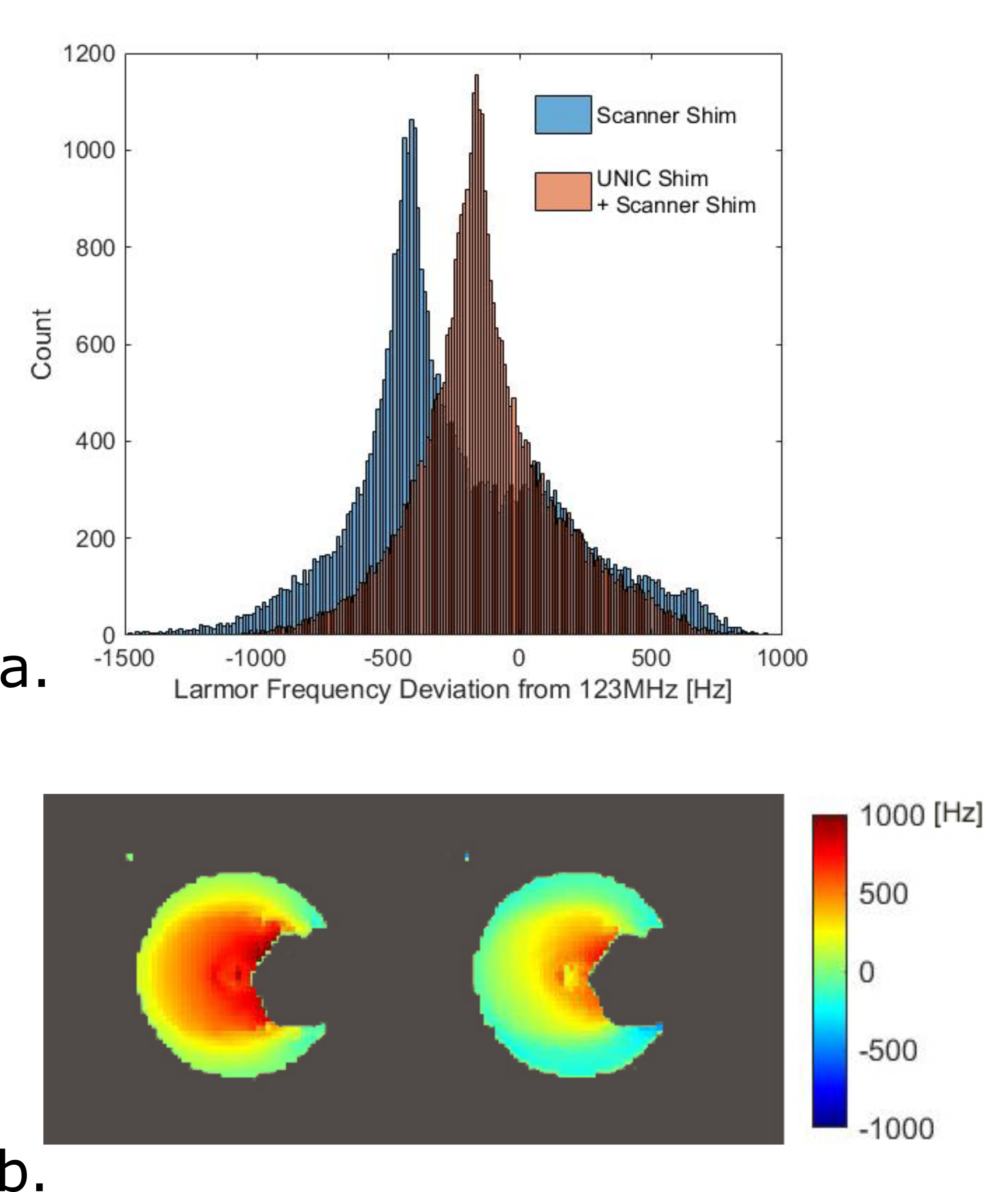
UNIC shimming reduced the opaque volume around the metal prosthesis (Figures 3,4). For TE=2.49ms, in all but one of the 29 slices in the 3D-shimmed volume, UNIC reduced signal void and revealed more of the area around the prosthesis. For TE=4.80ms, UNIC reduced signal void in 21 slices, but increased it in the remaining 8. Computed from signal and void area measurements and slice thickness, the opaque volume around the prosthesis reclaimed and made visible was approximately 24.0cm3 +/- 0.5cm3 at TE=2.49ms, and 22.5cm3 +/- 0.5cm3 at TE=4.8ms. Some of the best improvement was at slices across the femoral head (Figures 3a,b and 4), where some common complications of hip arthroplasty originate, including particle-mediated osteolysis [19, 20]. UNIC shimming reduced the dispersion of and shifted closer to 0Hz the off-resonance Larmor frequency distribution (Figure 5), indicating improved overall B0 homogeneity in the shimmed volume.Conclusion
The unified shim-RF coil array can improve B0 homogeneity around metal over the scanner shim alone. We observed reduction in the metal-induced signal void artifact in a hip prosthesis phantom using GRE scans, generally more susceptible than spin echo scans to metal artifacts. There is a potential that the sequence-agnostic unified shim-RF array technology will enable clinicians to see and assess tissue confidently closer to metal, including prostheses and interventional devices, including with the generally faster GRE, currently largely excluded in clinic for near-metal scanning. Further studies and in-vivo validations are under way.Acknowledgements
Authors extend their gratitude to Drs. Bernd Stoeckel, Fraser Robb, and Miguel Navarro. This work was supported by NIH U01 EB028145; R01 HL156818.References
1. Lu, W., et al., SEMAC: Slice Encoding for Metal Artifact Correction in MRI. Magn Reson Med, 2009. 62(1): p. 66-76.
2. Choi, S.J., et al., Metal artifact reduction with MAVRIC SL at 3-T MRI in patients with hip arthroplasty. AJR Am J Roentgenol, 2015. 204(1): p. 140-7.
3. Cho, Z.H., D.J. Kim, and Y.K. Kim, Total inhomogeneity correction including chemical shifts and susceptibility by view angle tilting. Med Phys, 1988. 15(1): p. 7-11.
4. Sutter, R., et al., Reduction of metal artifacts in patients with total hip arthroplasty with slice-encoding metal artifact correction and view-angle tilting MR imaging. Radiology, 2012. 265(1): p. 204-14.
5. Olsen, R.V., et al., Metal artifact reduction sequence: early clinical applications. Radiographics, 2000. 20(3): p. 699-712.
6. Juchem, C., et al., Dynamic multi-coil shimming of the human brain at 7 T. J Magn Reson, 2011. 212(2): p. 280-8.
7. Han, H., A.W. Song, and T.K. Truong, Integrated parallel reception, excitation, and shimming (iPRES). Magn Reson Med, 2013. 70(1): p. 241-7.
8. Truong, T.K., D. Darnell, and A.W. Song, Integrated RF/shim coil array for parallel reception and localized B0 shimming in the human brain. Neuroimage, 2014. 103: p. 235-40.
9. Darnell, D., T.K. Truong, and A.W. Song, Integrated parallel reception, excitation, and shimming (iPRES) with multiple shim loops per radio-frequency coil element for improved B0 shimming. Magn Reson Med, 2017. 77(5): p. 2077-2086.
10. Stockmann, J.P., et al., A 32-channel combined RF and B0 shim array for 3T brain imaging. Magn Reson Med, 2016. 75(1): p. 441-51.
11. Talbot, B.S. and E.P. Weinberg, MR Imaging with Metal-suppression Sequences for Evaluation of Total Joint Arthroplasty. Radiographics, 2016. 36(1): p. 209-25.
12. Fritz, J., et al., MR imaging of hip arthroplasty implants. Radiographics, 2014. 34(4): p. E106-32.
13. Fritz, J., B. Lurie, and H.G. Potter, MR Imaging of Knee Arthroplasty Implants. Radiographics, 2015. 35(5): p. 1483-501.
14. Han, H., Multi-Coil B0 Shimming in the Body, Program E0912, in Educational Proceeding of ISMRM and SMRT Annual Meeting and Exhibition, Paris, France, Virtual. 2020.
15. Han H, S.J., Cao W, Li Z, Cho J, Zhou D, Wang Y, and Li D, Unified Coils (UNIC) for Parallel Imaging and B0 Shimming., in European Society for Magnetic Resonance in Medicine and Biology (ESMRMB) 33rd Annual Scientific Meeting. 2016: Vienna, Austria.
16. Yang HJ, S.J., Azab L, Liu W, Lu M, Huang Y, Yoosefian G, Selvin S, Handelin R, Shan Y, Serry FM, Xie Y, Christodoulou AG, Bi X, Dharmakumar R, Li D, and Han H. High-Order B0 Shimming at 3T Using a UNIfied Coil (UNIC) for RF receive and shimming (program 2183). in ISMRM and SMRT Annual Meeting and Exhibition. 2020. Paris (Virtual).
17. MathWork, N., MA, USA, Matlab. 2020.
18. Rasband, W., ImageJ. U. S. National Institutes of Health: Bethesda, Maryland, USA.
19. Goodman, S.B. and J. Gallo, Periprosthetic Osteolysis: Mechanisms, Prevention and Treatment. J Clin Med, 2019. 8(12).
20. Pajarinen, J., et al., Particle disease really does exist. Acta Orthop, 2018. 89(1): p. 133-136.
Figures