3105
In-vivo B0 shimming of the liver using a local array of shim coils in combination with 2nd order spherical harmonics at 7T1Radiology Department, UMC Utrecht, Utrecht, Netherlands, 2MR Shim GmbH, Reutlingen, Germany
Synopsis
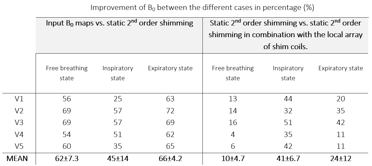
Maximizing shim performance, particularly for larger organs such as the liver, is crucial at ultra-high field MRI due to the increased sensitivity to B0 field inhomogeneity. The conventional shimming in most MRI systems at ultra-high field, provides insufficient correction. However, by combining the available 2nd order spherical harmonic fields with an external array of 16 local shim coils, the magnetic field homogeneity in the liver is improved by as much as 44%.
Introduction
The increase in SNR and CNR at ultra-high field MRI (7T) allows for higher spatial resolutions compared to lower magnetic fields, which makes ultra-high field MRI a tool with strong clinical potential. However, static inhomogeneity of the main magnetic field (B0) throughout the human body leads to local frequency offsets that result in artefacts during imaging (MRI) and spectroscopic measurements.[1] To compensate for this, it has been shown that higher order spherical harmonics (SH) are needed in addition to the conventional B0 shimming methods of the scanner.[2] Alternatively, local shim coil arrays that provide additional degrees of freedom albeit confined to a small region, can be used for a high number of shim coils and channels in the MR scanner.[3] In addition, it is possible to generate spatially varying magnetic fields that counteract the B0 field variations in real time, since the local shim coils couple less to the conductive bore (shim coils) reducing eddy currents. In previous works of Van den Wildenberg et al., it was demonstrated that the B0 field variations in simulations can be substantially reduced by using a local array of shim coils compared to the standard hardware in the liver.[4, 5] The aim of this work is to further improve B0 field homogeneity in the liver in-vivo by applying second order SH fields in combination with an array of 16 independently controlled external shim coils.Methods
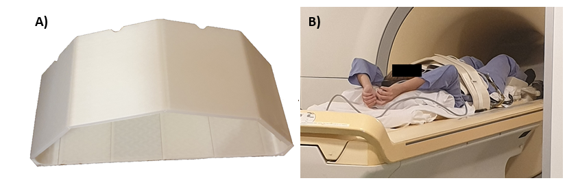
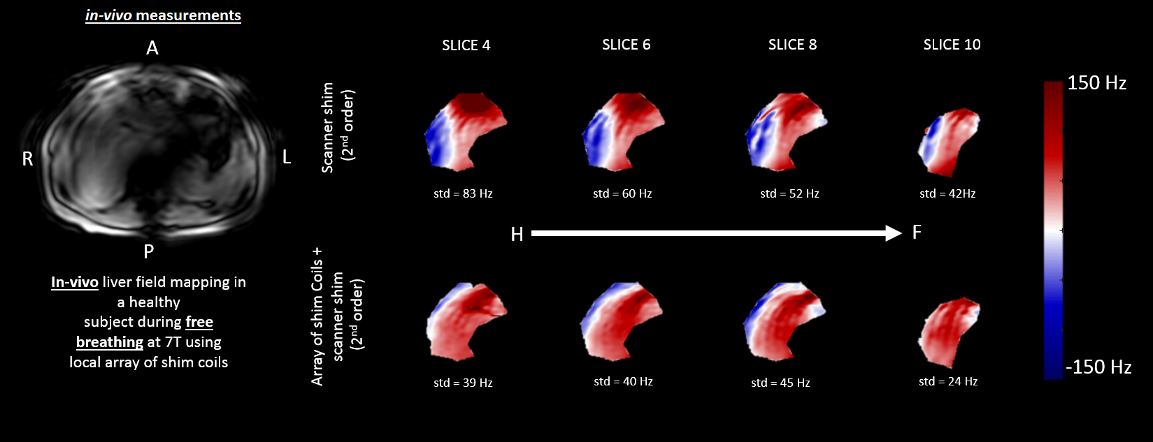
Simulations were done in previous works using a local array of shim coils. [4, 5] A whole-body 7T MR scanner equipped with a multi-transmit RF system (Achieva, Philips Healthcare, Best, the Netherlands) was used to acquire B0 maps of the liver in a healthy volunteer. Eight transceiver fractionated dipole antennas with 16 additional receive loops interfaced to 8 parallel 2kW peak power amplifiers, were positioned symmetrically around the body at the position of the liver.[6] 16 circular enameled copper local shim coils (each with a diameter of 7.5cm and 14turns) and Jupiter shim amplifiers for driving the shim coils were provided by MR Shim GmbH (Reutlingen, Germany). The array of shim coils was positioned around the body array and was arranged in two uniform rows of four, eight on the bottom and eight on top, see Figure 1. In one healthy volunteer, seventeen dual-echo B0 maps (GE, 282×402×78mm3 FOV, 6×6×6mm3 voxel size, FA=5°, TR=10ms, TE=1.493ms, ΔTE=1ms) were acquired during free breathing. Sixteen of the seventeen B0 maps were used as calibration data for the array of shim coils to gather channel-specific B0 maps (each channel had a current of 1.5 Amps). A 2nd order shimmed B0 map was acquired as a reference. The optimal current for each shim coil was calculated to minimize the least-squares deviation of the total field (∆B0(x,y,z) + shim coil created field) in the region of interest using the tool Arche (MR Shim GmbH, Reutlingen, Germany). Two types of B0 maps were measured for method comparison: 1) second order shimming using the standard hardware in the scanner and 2) second order shimming in combination with the array of shim coils. The standard deviation of both shimmed fields in the region of interest were calculated and expressed in Hertz (γB0). The percentage change in standard deviation was calculated and used as a measure for the improvement of shim performance.Results
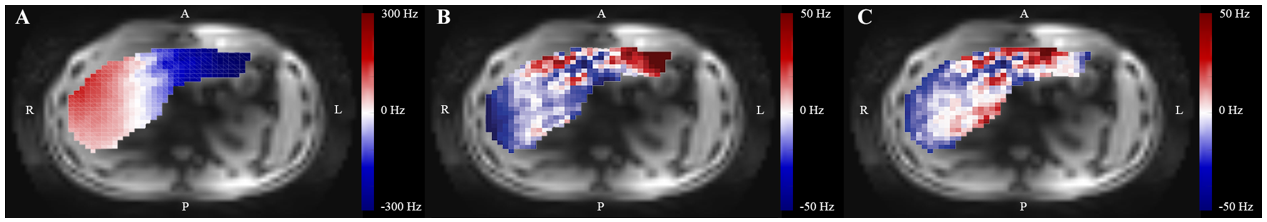
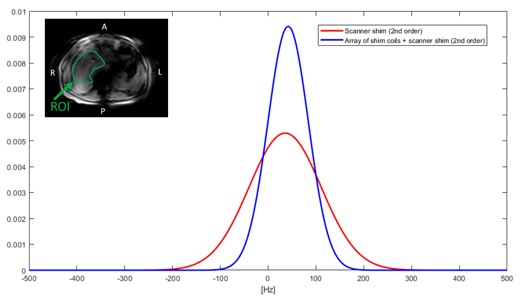
Previous simulation results are shown in Figure 2 and 3. [4, 5] The currents for the in-vivo measurements for each shim coil calculated in Arche are in the range of -0.6A and +0.4A. The standard deviation of the B0 field in the region of interest of the B0 map using the second order shimming is 75.4Hz, When combined with the array of shim coils this value reduces to 42.4Hz (Figure 4), representing a 44% improvement in shimming performance. The B0 field throughout the liver in the transversal plane from four imaging slices is shown in Figure 5.Discussion
Substantial improvement in field uniformity can be obtained using an additional local array of shim coils for a specific organ when compared to only the standard second order B0 shimming. In our study, we measured B0 maps during free breathing, because real-time shim updating is not implemented yet. Future studies will focus on combining our previous work which showed improved shim fields by shim updating during the different phases of respiratory motion and this work.[4] The 44% B0 homogeneity improvement measured is much better than the 10% improvement that was obtained in simulation (Figure 2/3).[4] The discrepancy can at least partially be explained by a different BMI of the volunteer in comparison with the simulations, size of the liver, and the fact that larger shim coils were used in practice (the shim coil geometry between simulations and the in-vivo measurements are slightly different, d=5cm & 20turns, d=7.5cm & 16turns respectively).Conclusion
This study shows that local B0 field variations in-vivo at ultra-high-field can be reduced using a local array of shim coils by 44%. As local shimming becomes more important at ultra-high magnetic field strengths, the use of local shim coil arrays will become a necessity, particularly for larger organs such as the liver.Acknowledgements
No acknowledgement found.References
[1] T. K. Truong, D. W. Chakeres, D. Q. Beversdorf, D. W. Scharre, and P. Schmalbrock, “Effects of static and radiofrequency magnetic field inhomogeneity in ultra-high field magnetic resonance imaging,” Magnetic Resonance Imaging, vol. 24, no. 2. pp. 103–112, 2006, doi: 10.1016/j.mri.2005.09.013.
[2] J. W. Pan, K. M. Lo, and H. P. Hetherington, “Role of very high order and degree B0 shimming for spectroscopic imaging of the human brain at 7 tesla,” Magnetic Resonance in Medicine, vol. 68, no. 4. pp. 1007–1017, 2012, doi: 10.1002/mrm.24122.
[3] C. Juchem and R. A. de Graaf, “B0 magnetic field homogeneity and shimming for in vivo magnetic resonance spectroscopy,” Anal. Biochem., pp. 1–13, 2016.
[4] L. van den Wildenberg et al., “B0 shimming of the liver using a local array of shim coils in the presence of respiratory motion at 7T,” in ISMRM 27th annual meeting, 2019, p. #0221.
[5] L. van den Wildenberg et al., “B0 shimming of the liver using a local shim coil array at ultra-high-field MRI,” in ISMRM 28th annual meeting, 2020, p. #4220.
[6] B. R. Steensma et al., “An 8-channel Tx Rx dipole array combined with 16 Rx loops for high-resolution functional cardiac imaging at 7 T,” Magn. Reson. Mater. Physics, Biol. Med., vol. 31, pp. 7–18, 2018.
Figures