3034
Simultaneous multi-parameter mapping to characterize Parkinson’s Disease using strategically acquired gradient echo imaging1Department of Medical Imaging, Henan Provincial People’s Hospital & Zhengzhou University, Zhengzhou, China, 2MR Collaboration, Siemens Healthcare Ltd. Beijing China, Beijing, China, 3Wayne State University, Detroit, MI, United States, 4Shanghai Zhuyan Medical Technology Company, Shanghai, China
Synopsis
Scanning patients with Parkinson’s disease (PD) is challenging because of tremors. Strategically acquired gradient echo (STAGE) imaging can acquire seven quantitative images relatively quickly. This method could provide comprehensive evaluations of cerebral alterations in PD patients while reducing motion artifacts. This study compared T1, T2*, R2*, and susceptibility-weighted imaging mapping (SWIM) acquired with STAGE imaging between 15 PD patients and 15 healthy controls (HC). Significant differences were found for the T1 and T2* values in several brain areas between PD and HC, suggesting that STAGE imaging is a convenient and powerful tool to investigate PD.
Introduction
Parkinson’s disease (PD) is a common neurodegenerative disease. The function of some brain regions in PD patients is selectively impaired. PD patient tremors make magnetic resonance imaging (MRI) challenging causing several motion artifacts. Therefore, shortening MRI times is essential for acquiring high-quality multimodal imaging for PD analyses. Strategically acquired gradient echo (STAGE) can provide nine qualitative images and seven quantitative images in 5 minutes or less at 3T [1,2,3]. STAGE imaging resulted in shorter scan time with greater amounts of information compared to other single-modal imaging. This multimodal imaging technique with whole-brain analyses was found to be more convenient and accurate for use in PD patients. In this study, we acquired T1, T2*, R2*, and susceptibility-weighted imaging mapping (SWIM) using STAGE imaging to analyze the difference between PD patients and healthy controls.Methods
STAGE data of 15 PD patients (six women and nine men, age: 59.5 years ± 5.8 years) and 15 healthy controls (nine women and six men, age: 58.9 years ± 4.7 years) were acquired on a 3T MAGNETOM Prisma scanner (Siemens Healthcare, Erlangen, Germany) equipped with a 64-channel head/neck coil. The protocol included two 3D gradient echo (GRE) scans for STAGE and T1 MPRAGE sequences. The parameters of two GRE scans for STAGE were: flip angle1=6°; flip angle2 = 24°, TR =25ms, TE1=7.5/17.5 ms for two echoes; TE2 = 8.75/18.75ms for two echoes; voxel size=0.7 × 0.7 × 2 mm3, matrix size=288 × 384 × 64, FOV= 256 × 256mm2. The total scan time was about 5 minutes. T1-weighted enhanced (T1-WE), T1, T2*, R2*, and SWI maps provided with STAGE imaging were processed using SPIN (Signal Processing in NMR) software for whole-brain analyses. The T1WE images were specifically registered to T1 MPRAGE images using Statistical Parametric Mapping 12 (SPM12; https://www.fil.ion.ucl.ac.uk/spm/software/spm12/). The deformation field generated from T1 MPRAGE image segmentation was applied to the T1WE, T1, T2* and R2* images. The differences in the images created by T1, T2*, R2*, and SWI mapping between PD patients and healthy controls were analyzed by assessing each region of interest (ROI), including 32 sub-cortical regions and 96 cortical regions using masks generated from the Harvard-Oxford atlas. An independent samples t-test was conducted using SPSS 22.0 software (IBM SPSS Inc., Chicago, IL, USA), and a P of <0.05 was considered statistically significant.Results
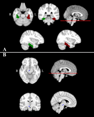
T1-weighted enhanced (T1-WE), T1, T2*, R2*, and SWI maps were provided with STAGE imaging (as shown in Figure 1). For sub-cortical brain areas, R2* of right parabrachial pigmented nucleus (PBP) was significantly higher in PD patients than in healthy controls (p <0.05). T2* and T1 mapping of the right PBP was significantly lower in PD patients than in healthy controls (p <0.05), as shown in Table 1, figure 2. For cortical brain areas, R2* of the right temporal fusiform cortex, posterior division (TFCp) was significantly higher in PD patients than in healthy controls. T2* mapping of the left and right TFCp was significantly lower in PD patients than in healthy controls, as shown in Table 2 and figure 2.Discussion
PD is one of the most common neurodegenerative disorders, and the pathologic signs include a loss of dopaminergic neurons with Lewy body formation. Elevated iron levels can be deduced from MRI with decreasing T2* signals and increasing R2* signals [4]. One of the main iron-accumulating regions in PD patients is the brainstem[5], which is close to the PBP. Previous studies have found PD patients with significant iron accumulation in the fusiform cortex [5], which could explain our findings that R2* signals were significantly higher in the temporal fusiform cortex of PD patients. The STAGE differences seen between PD patients and healthy controls explain the pathologic differences observed. Remaining still is a problem for PD patients undergoing MRI. STAGE imaging provides 16 different qualitative images in 10 minutes resulting in similar accuracy and better quality compared to conventional imaging, helping to resolve movement artifacts.Conclusion
STAGE is a fast and accurate quantitative imaging method that can sensitively detect subtle structural brain changes and iron accumulation in patients with PD.Acknowledgements
National Key R&D Program of China (2017YFE0103600), National Natural Science Foundation of China (81720108021), Zhongyuan Thousand Talents Plan Project(ZYQR201810117), Zhengzhou Collaborative Innovation Major Project (20XTZX05015)References
[1] Chen Y , Liu S , Wang Y , et al. STrategically Acquired Gradient Echo (STAGE) imaging, part I: Creating enhanced T1 contrast and standardized susceptibility weighted imaging and quantitative susceptibility mapping[J]. Magnetic Resonance Imaging, 2017:S0730725X1730228X.
[2] Wang Y , Chen Y , Wu D , et al. STrategically Acquired Gradient Echo (STAGE) imaging, part II: Correcting for RF inhomogeneities in estimating T1 and proton density[J]. Magnetic Resonance Imaging, 2018:S0730725X17302291.
[3] C, E. Mark Haacke A B , et al. STrategically Acquired Gradient Echo (STAGE) imaging, part III: Technical advances and clinical applications of a rapid multi-contrast multi-parametric brain imaging method - ScienceDirect[J]. Magnetic Resonance Imaging, 2020, 65:15-26.
[4] Ulla M , Bonny J M , Ouchchane L , et al. Is R2* a New MRI Biomarker for the Progression of Parkinson's Disease? A Longitudinal Follow-Up[J]. Plos One, 2013, 8(3):e57904.
[5] Julio A C , Arturo C B , Betts M J , et al. The whole-brain pattern of magnetic susceptibility perturbations in Parkinson's disease[J]. Brain(1):118-131.
Figures


