3033
Cerebral hyper-perfusion associated with mild cognitive impairment in de novo Parkinson’s disease1GE Healthcare, Shanghai, China, 2Zhongshan Hospital, Shanghai, China, 3GE Healthcare, Beijing, China
Synopsis
We aimed to investigate the patterns of cerebral perfusion changes in early drug-naive PD patients with mild cognitive impairment (PD-MCI). Voxel-wise cerebral blood flow (CBF) were compared among PD-MCI patients, PD patients with normal cognition (PD-NC) and healthy controls. Increased CBF was detected in the right anterior cingulate cortex in the PD-MCI group as compared to the controls, as well as higher perfusion in the right middle frontal gyrus and superior frontal gyrus relative to PD-NC. Cerebral hyper-perfusion in frontal lobe might be associated with cognitive decline in early de novo PD.
Purpose
Parkinson's disease (PD) is the second neurodegenerative disease in the elderly, manifested as classic motor symptoms and non-motor symptoms. Cognitive impairment is one of the well-defined non-motor symptoms and seriously affects the quality of life of patients with PD. The mechanism underlying cognitive impairment in PD is still unclear. Brain structural and functional abnormalities, including volume loss, cortical thinning and cerebral perfusion alterations, were reported associated with PD patients with mild cognitive impairment (MCI). However, these findings were not consistent. Recently, three-dimensional pulsed continuous arterial spin labeling (3D pCASL) was considered an advanced method for cerebral blood flow (CBF) measurement.1 Voxel-based analysis (VBA) were used to observe the regional CBF which could improve the accuracy of CBF quantification.2 In this study, we aimed to investigate the different patterns of cerebral perfusion changes in the early drug-naïve PD-MCI patients as compared with the PD patients with normal condition (PD-NC) and normal controls using 3D pCASL imaging with voxel-based analysis.Methods
All the subjects gave written informed consent to participate the study, which was approved by the local ethical committee. The newly diagnosed and drug naïve PD patients (n=63) were recruited from the movement disorders clinic at Zhongshan hospital. The diagnosis of PD was made according to the Movement Disorder Society (MDS) clinical diagnostic criteria for Parkinson's disease, and the diagnosis of PD-MCI (n=20) was according to the level 2 criteria recommended by the MDS Task Force 2012. The patients who did not meet the criteria for PD-MCI were classified as PD-NC (n=43). All age- and gender- matched control subjects (n=42) were voluntarily recruited from the community and had no history of neurologic or psychiatric disorders. Perfusion scans were performed with a 3.0-T MR750 scanner (GE Healthcare, Milwaukee, WI) using an 8-channel phase array head coil. Two sets of ASL images were acquired with and without the spatially selective inversion (tagging) pulse (TR/TE 4830/10 ms, flip angle 155°, labeling duration 1.5 s, post label delay 1.5 s, matrix =128x128, FOV 24 cm, thickness/gap 4/0 mm). The difference maps between the tag and control pairs were averaged for each subject and quantitative cerebral blood flow (CBF) maps were calculated with the vender provided toolbox. Image pre-processing was performed using SPM12 (http://www.fil.ion.ucl.ac.uk/spm). Spatial transformation included a three-dimensional rigid body registration to correct for head motion, followed by a nonlinear warping to spatially normalized CBF maps into a standard stereotaxic space. The normalized CBF maps were resampled to 2x2x2 mm3 isotropic voxel size and smoothed with a 6 mm isotropic Gaussian kernel. Voxel-based analyses was conducted using one-way ANOVA to make comparison among PD-MCI, PD-NC and control groups. The post-hoc two-sample T test was used to compare the differences between each two groups. The cluster-level Family Wise Error (FWEc) correction implemented in SPM12 was used for multiple comparison correction (corrected p<0.05).Results
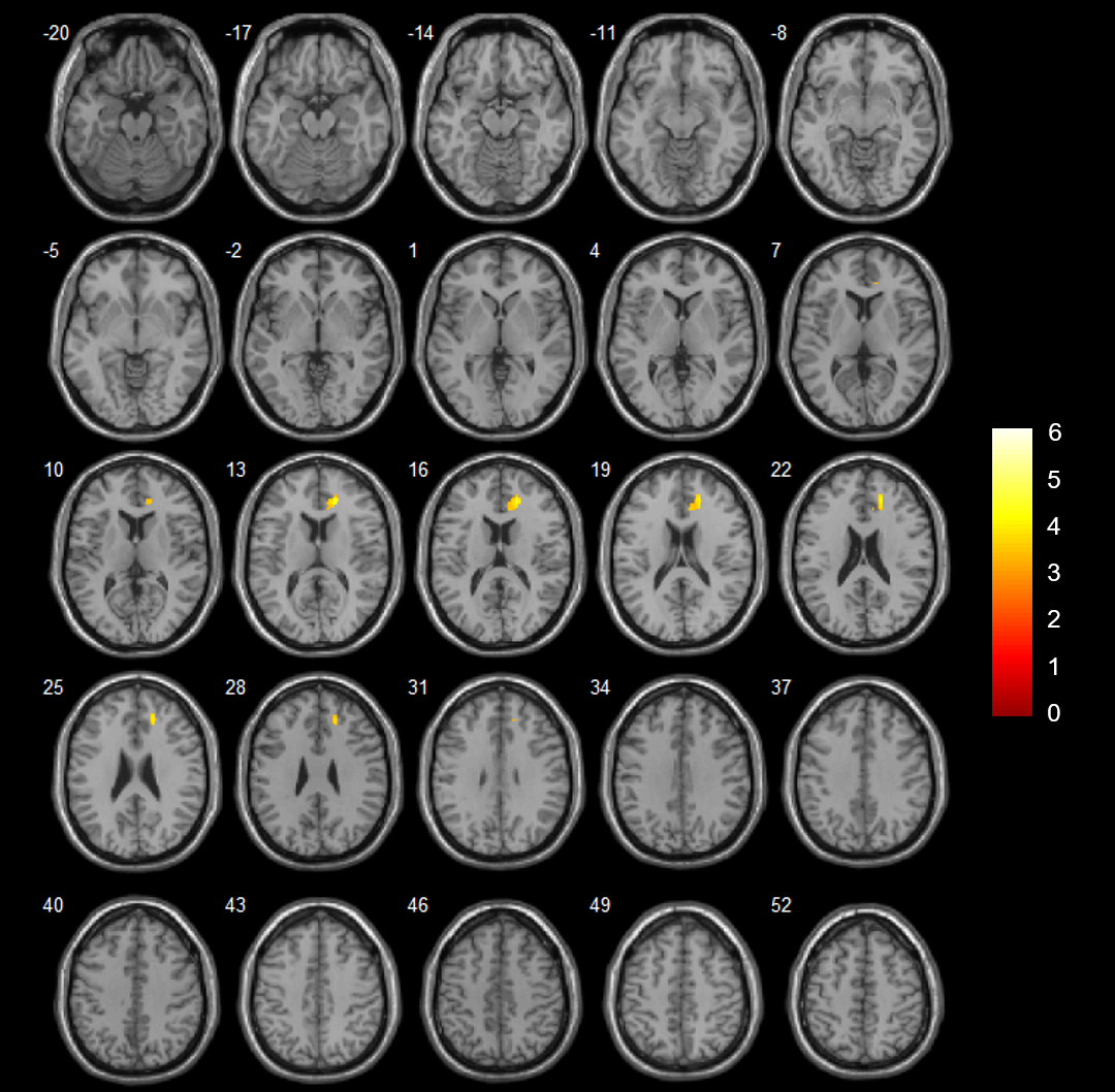
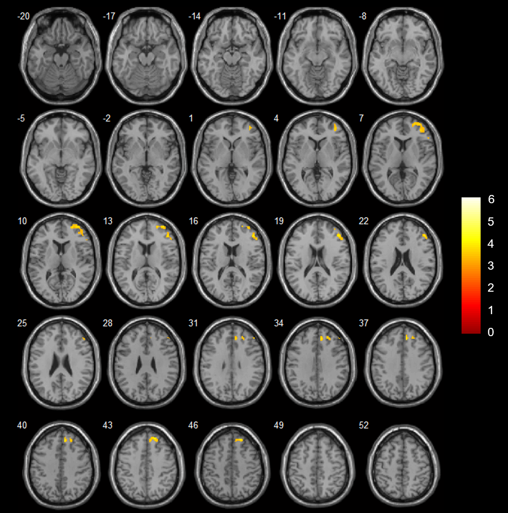
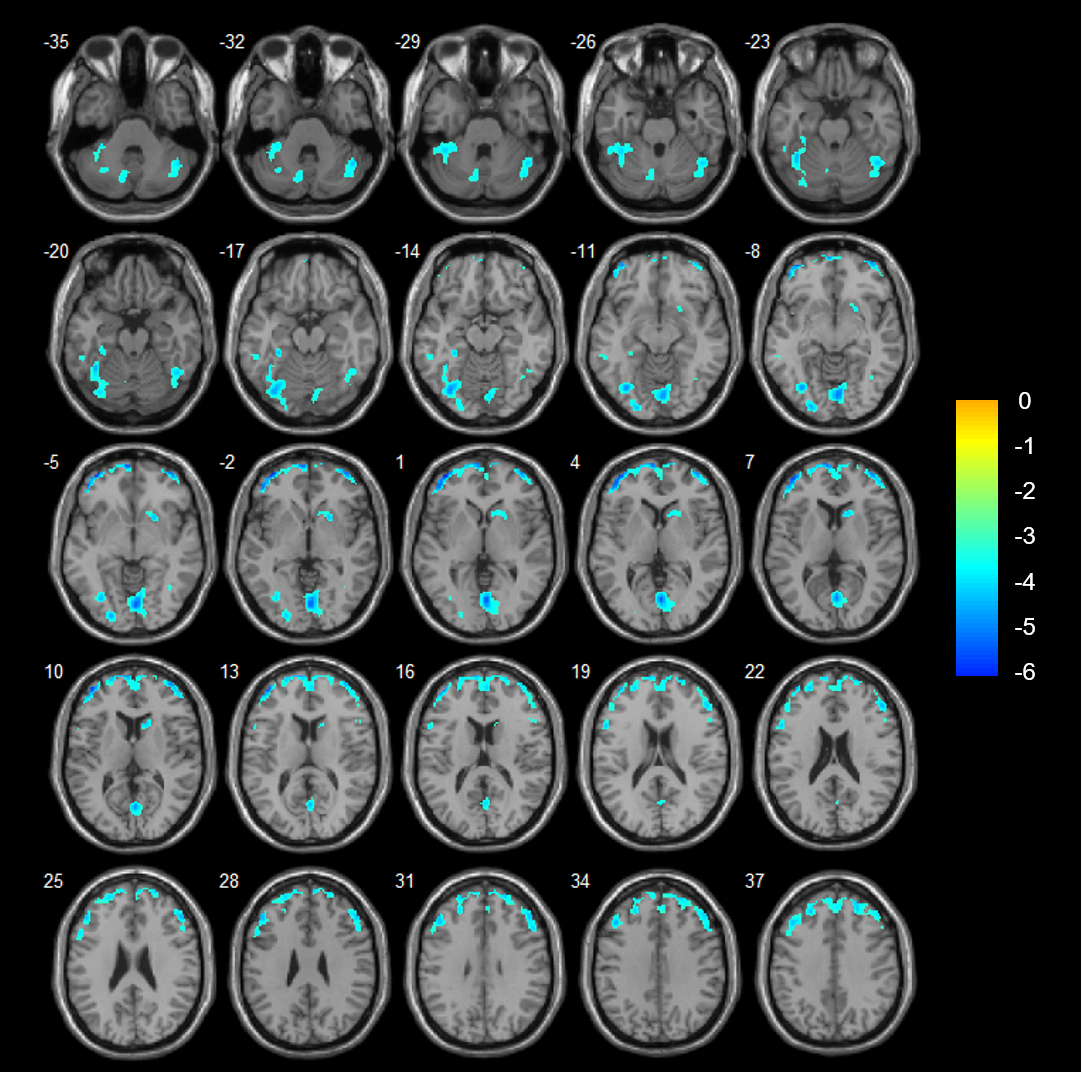
PD-MCI showed significantly increased CBF values in the right anterior cingulate cortex (ACC) as compared with controls (Fig. 1). In addition, PD-MCI exhibited significantly higher perfusion in the right MFG and right SFG than those in PD-NC (Fig. 2). However, compared with controls, significantly decreased CBF was detected in the left MOG, right lingual gyrus, left MFG, right sub-lobar region (mainly caudate and putamen) and bilateral cerebellum posterior lobe in the patients with PD-NC (Fig. 3).Discussion and Conclusion
The present study aimed to investigate the patterns of cerebral perfusion changes in early de novo PD with different cognitive states. By examining only drug-naïve patients in our study, the potential confounding effects of medication or long disease progression were eliminated. Our results revealed increased perfusion in the right middle frontal gyrus, right superior frontal gyrus and right anterior cingulate in PD-MCI patients as compared to PD-NC. In addition, PD-MCI showed increased CBF values in the right ACC as compared to normal controls. We suggested that the hyper-perfusion in the frontal lobe and limbic lobe might reflect an adaptive compensatory mechanism in response to cognitive decline in early PD-MCI.Acknowledgements
No acknowledgement found.References
1. Dai W, Garcia D, de Bazelaire C, et al. Continuous flow-driven inversion for arterial spin labeling using pulsed radio frequency and gradient fields. Magn Reson Med. 2008;60(6):1488-1497.
2. Huang D, Wu B, Shi K, et al. Reliability of three-dimensional pseudo-continuous arterial spin labeling MR imaging for measuring visual cortex perfusion on two 3T scanners. PLoS One. 2013;8 (11): e79471.
Figures