3030
Value of magnetic resonance fingerprinting for differentiating tremor-dominant Parkinson’s disease from essential tremors1Department of Radiology, Zhengzhou University People’s Hospital & Henan Provincial People’s Hospital, Academy of Medical Sciences., Zhengzhou, China, 2Henan Key Laboratory for Medical Imaging of Neurological Diseases, Zhengzhou, Henan, China, ZhengZhou, China, 3Department of Radiology, Henan University People’s Hospital & Henan Provincial People’s Hospital, School of Basic Medicine., Zhengzhou, China, 4MR Collaboration, Siemens Healthcare Ltd., Beijing, China, BeiJing, China, 5MR Pre-development, Siemens Healthcare GmbH, Erlangen, Germany, Erlangen, Germany
Synopsis
Conventional magnetic resonance imaging cannot reliably differentiate Parkinson’s disease (PD) from essential tremors (ET). Magnetic resonance fingerprinting (MRF) can simultaneously acquire T1 and T2 relaxometry. This study utilized MRF to obtain T1 and T2 values in substantia nigra (SN) of patients with tremor-dominant PD and ET. The T1 values of SN were significantly higher in patients with tremor-dominant PD than those with ET, whereas the T2 values showed no significant differences between groups. The findings suggest that MRF T1 mapping of the SN can potentially differentiate tremor-dominant PD from ET
Introduction
Parkinson’s disease (PD) is a common movement disorder characterized by dopamine neuronal loss in the substantia nigra (SN) on histopathology. However, tremor-dominant PD is frequently misdiagnosed as essential tremors (ET). Quantitative T1 and T2 maps can be acquired using conventional magnetic resonance imaging (MRI) relaxometry methods, which need relatively long scan times and have not been used to differentiate PD from ET. MR fingerprinting (MRF) can simultaneously acquire T1 and T2 maps, significantly shortening acquisition times and yielding perfectly aligned images compared to conventional MRI relaxometry methods.[1,2] The purpose of this study was to assess MRF values to differentially diagnose tremor-dominant PD from ET.Methods
A total of 10 patients (mean age: 59 years; 6 males and 4 females) [BM(DMM1] with tremor-dominant PD and 10 age- and sex-matched patients with ET were enrolled. Conventional MRI included T2-weighted and T1-weighted imaging, and MRF[KG(DMDM2] [1] data were acquired using a 3T MAGNETOM Skyra MRI scanner (Siemens Healthcare, Erlangen, Germany) with a 20-channel head/neck coil. A prototypic spiral fast imaging with steady-state free-precession MRF[2] [KG(DMDM3] with B1+ [KG(DMDM4] correction[3] was performed on all patients (field of view = 256 x 256 mm2; matrix = 256 x 256; slice thickness = 5 mm; flip angles = 0°– 74°; repetition time = 12.1 ms -15.0 ms; 3000 measurements; 41 s/slice; 5 slices). For each patient, regions of interest (ROIs) were manually drawn on the T2-weighted images to extract the bilateral SN. Then, the ROIs were copied to the MRF-derived T1 and T2 maps, respectively. The Mann-Whitney U test was used to evaluate the T1 and T2 value differences in the ipsilateral and contralateral SN between the two groups. P < 0.05 was considered statistically significant. [BM(DMM1]age, gender? [KG(DMDM2]Please cite the original MRF publication here. [KG(DMDM3]Please cite the method: https://onlinelibrary.wiley.com/doi/10.1002/mrm.25559 [KG(DMDM4]Please cite: https://www.ncbi.nlm.nih.gov/pmc/articles/PMC2929762/Results
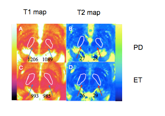
MRF T1 and T2 mapping results from two representative subjects were shown in figure 1. The mean MRF-derived T1 value of the bilateral SN was significantly higher in patients with tremor-dominant PD than those with ET (1010±37 ms vs 958±30 ms, P < 0.001). The mean MRF-derived T2 value of the bilateral SN was not significantly different between the two groups (P > 0.05).Discussion
Dopamine neuronal loss and neuroinflammation related to degeneration could lead to increased free water content in the SN of patients with PD. MRF-derived T1 and T2 values were increased with elevated water content. The T1 value in the SN was significantly higher in patients with tremor-dominant PD than in those with ET, whereas the T2 value could not differentiate PD from ET. This result might be caused by the higher iron content in the SN of patients with PD. Although iron content decreases T1 and T2 values, iron content influences T2 values more frequently than T1 values.Conclusion
MRF T1 mapping in the SN might be able to differentiate tremor-dominant PD from ET and potentially guide treatment strategies.Summary of Main Findings
The use of magnetic resonance fingerprinting revealed that the substantia nigra T1 values were significantly higher in patients with tremor-dominant Parkinson’s disease than those with essential tremors.Acknowledgements
National Key R&D Program of China (2017YFE0103600), National Natural Science Foundation of China (81720108021), Zhongyuan Thousand Talents Plan Project(ZYQR201810117), Zhengzhou Collaborative Innovation Major Project (20XTZX05015)References
1. Jiang Y, Ma D, Keenan KE, Stupic, KF, Gulani V, Griswold MA (2017) Repeatability of Magnetic Resonance Fingerprinting T1 and T2 Estimates Assessed Using the ISMRM/NIST MRI System Phantom. Magn Reson 78:1452-1457.
2. European Society of Radiology (ESR) (2015) Magnetic Resonance Fingerprinting - a promising new approach to obtain standardized imaging biomarkers from MRI. Insights Imaging 6:163-165.
3. Jiang Y, Ma D, Seiberlich S, Gulani V, Griswold MA (2015). MR fingerprinting using fast imaging with steady state precession (FISP) with spiral readout 74:1621-1631.
4. Chung S, Kim D, Breton E, Axel L (2010). Rapid B1+ Mapping Using a Pre-Conditioning RF Pulse with TurboFLASH readout. Magn Reson Med 64(2): 439–446.
Figures