3022
Substantia nigra magnetic resonance spectrum in differentiating tremor-dominant Parkinson’s disease from essential tremors
Rushi Chen1, Yan Bai1, Qin Feng1, Menghuan Zhang1, Xianchang Zhang2, and Meiyun Wang1
1Henan provincial people's hospital, Zhengzhou, China, 2MR Collaboration, Siemens Healthcare Ltd, Beijing, China, Beijing, China
1Henan provincial people's hospital, Zhengzhou, China, 2MR Collaboration, Siemens Healthcare Ltd, Beijing, China, Beijing, China
Synopsis
Conventional magnetic resonance imaging has limitations in differentiating Parkinson’s disease (PD) from essential tremors (ET). Magnetic resonance spectroscopy (MRS) can non-invasively detect neurochemical alterations in biological tissues. In this study, we used MRS to obtain N-acetylaspartate (NAA)/creatine (Cr) and choline (Cho)/Cr ratios in the substantia nigra (SN) of patients with tremor-dominant PD and ET. The NAA/Cr ratio in the contralateral SN was significantly higher in patients with tremor-dominant PD than those with ET, whereas the Cho/Cr ratios showed no significant differences between groups. The findings suggest that MRS in the SN may be helpful in differentiating tremor-dominant PD from ET.
Introduction
Parkinson’s disease (PD) is a common movement disorder characterized by dopamine neuronal loss and neuroinflammation in the substantia nigra (SN) on histopathology. However, tremor-dominant PD is frequently misdiagnosed as essential tremors (ET). Magnetic resonance spectroscopy (MRS) can non-invasively detect neurochemical alterations such as N-acetylaspartate (NAA), creatine (Cr) and choline (Cho) in biological tissues. The purpose of this study was to assess NAA/Cr and Cho/Cr ratios derived from MRS to differentiate tremor-dominant PD from ET.Methods
A total of 12 patients (mean age: 62 years; 5 males and 7 females) with tremor-dominant PD and 12 age- and sex-matched patients with ET were enrolled in this study. High-resolution 3D T2-weighed image and signal voxel proton MRS data were collected on MAGNETOM Prisma 3T MR Scanner (Siemens Healthcare, Erlangen, Germany) with a 64-channel head-neck coil. A T2-SPACE sequence (field of view = 256 x 256 mm2; slice thickness = 1 mm; repetition time = 3200 ms; echo time = 407 ms) was performed on all patients. The 3D T2-weighted images were rebuilt in all three dimensions for SN. Single-voxel MRS (repetition time = 2000 ms; echo time = 30 ms) was performed on the SN of patients. The 1×1×1 cm3 voxel was placed on the left and right SN, respectively. The Mann-Whitney U test was used to evaluate the NAA/Cr and Cho/Cr ratios in the ipsilateral and contralateral SN between the two groups. P < 0.05 was considered statistically significant.Results
MRS results from two representative subjects were shown in figure 1. The NAA/Cr ratio of the contralateral SN was significantly decreased in patients with tremor-dominant PD than in patients with ET (2.01±0.55 vs 2.45±0.89, P < 0.01). However, the NAA/Cr ratio of the ipsilateral SN, as well as Cho/Cr ratio of the bilateral SN had no significant differences between the two groups (all P > 0.05).Discussion
Dopamine neuronal loss and neuroinflammation were the pathological characteristics in SN of PD. The NAA/Cr ratio in the contralateral SN was significantly higher in patients with tremor-dominant PD than in those with ET, whereas the NAA/Cr ratio of ipsilateral SN could not separate tremor-dominant PD from ET. This finding indicated the neuronal loss in the contralateral SN was more obvious compared with the ipsilateral SN in PD. In addition, the Cho/Cr ratio had no significant differences in the bilateral SN between tremor-dominant PD and ET. Cho/Cr was decreased with neuronal loss while increased with inflammatory cellular infiltration. Thus, the converse influence on Cho/Cr ratio of the SN may lead to no differences between tremor-dominant PD and ET.Conclusions
MRS in the SN may be a useful tool in differentiating tremor-dominant PD from ET.Acknowledgements
No acknowledgement found.References
Mazuel L, Chassain C, Jean B, Pereira B, Cladière A, Speziale C, Durif F. Proton Mr spectroscopy for Diagnosis and evaluation of Treatment efficacy in Parkinson Disease. Radiology. 2016;278(2):505-13.Figures
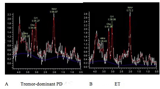
Figure 1. NAA/Cr was significantly decreased in the contralateral SN of a patient with tremor-dominant Parkinson’s disease (PD) (A) compared with that of a patient with essential tremors (ET).