3019
Locus coeruleus degeneration associated with less levodopa responsiveness in Parkinson’s Disease1Zhejiang University, Hangzhou, China
Synopsis
We conducted a neuromelanin sensitive magnetic resonance imaging, which is a good indicator for LC integrity, in 57 PD patients. We depicted a significant positive association between LC integrity and motor improvement after levodopa administration. We further confirmed this relationship in the level of objective brain alteration: LC integrity associated with the improvement of somatomotor network synchronization calculated from functional magnetic resonance imaging. We concluded that LC degeneration was an indicator for less levodopa responsiveness. LC integrity evaluation might be an alternative tool in predicting disease prognosis and stratifying patients into clinical trials for improving the efficacy of levodopa.
Introduction
Widely divergent responsiveness of levodopa in Parkinson’s disease (PD) patients is a vital clinical issue because of its relation with disease prognosis and therapeutic efficacy of deep brain stimulation. The positive association between the integrity of locus coeruleus (LC) noradrenergic system and levodopa responsiveness found in animal model has not been confirmed in vivo.Methods
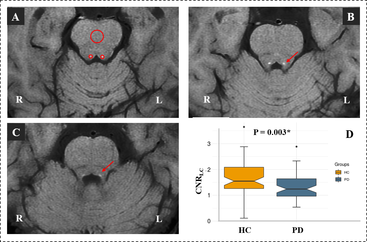
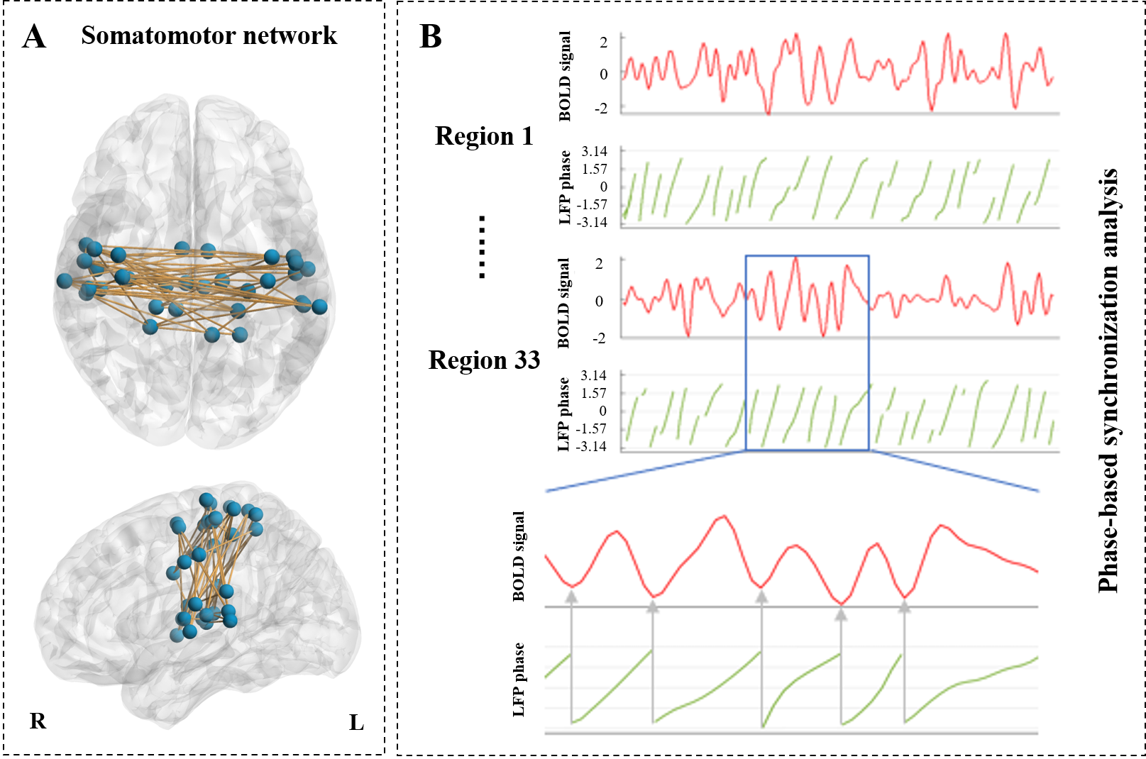
We conducted a neuromelanin sensitive magnetic resonance imaging, which is a good indicator for LC integrity, in 57 PD patients and 65 healthy controls (Figure 1). Levodopa challenge test was conducted for each patients. Then we evaluated the relationship between LC integrity and the change rate of Unified PD Rating Scale (UPDRS III) after levodopa administration. To further confirmed this association in the level of objective brain alteration, we assessed the relationship between LC integrity and the improvement of brain functional networks. A phase-based BOLD signal synchronization analysis was used to depict the organization of functional networks (Figure 2).Results
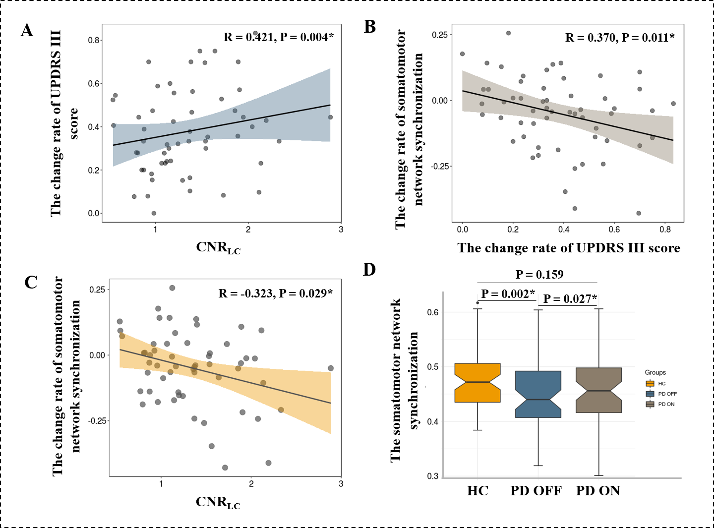
We depicted a significant positive association between LC integrity and motor improvement after levodopa administration (Figure 3A). The improvement of somatomotor network synchronization was associated with the improvement of UPDRS III (Figure 3B). We further confirmed that LC integrity associated with the improvement of somatomotor network synchronization (Figure 3C).In addition, PD patients showed decreased synchronization of somatomotor network during OFF state when comparing with HCs. And somatomotor network synchronization was significantly increased after levodopa administration (Figure 3D).
Conclusion
This study demonstrates that LC degeneration was an indicator for less levodopa responsiveness in PD. LC integrity evaluation using NM-MRI might be an alternative simple means in predicting disease prognosis and stratifying PD patients into clinical trials adding noradrenergic agents to dopaminergic medication.Acknowledgements
We thank all patients with Parkinson’s disease patients and healthy controls who participated in this study.References
1. De Pablo-Fernández E, Lees AJ, Holton JL, Warner TT. Prognosis and Neuropathologic Correlation of Clinical Subtypes of Parkinson Disease. JAMA neurology 2019;76:470-479.
2. Ostock CY, Lindenbach D, Goldenberg AA, Kampton E, Bishop C. Effects of noradrenergic denervation by anti-DBH-saporin on behavioral responsivity to L-DOPA in the hemi-parkinsonian rat. Behav Brain Res 2014;270:75-85.
3. Sulzer D, Cassidy C, Horga G, et al. Neuromelanin detection by magnetic resonance imaging (MRI) and its promise as a biomarker for Parkinson's disease. NPJ Parkinson's disease 2018;4:11.
4. Betts MJ, Kirilina E, Otaduy MCG, et al. Locus coeruleus imaging as a biomarker for noradrenergic dysfunction in neurodegenerative diseases. Brain 2019;142:2558-2571.
Figures