3015
Altered cortico-cerebellar functional connectivity of language processing in congenital blind children1Department of NMR & MRI Facility, All India Institute of Medical Sciences, Delhi, India, 2Dr RP Centre of Ophthalmology, All India Institute of Medical Sciences, Delhi, India
Synopsis
This study investigated structural alteration, cerebellar BOLD activation and functional connectivity during Braille reading by congenital blind (CB) children. Functional magnetic resonance imaging (fMRI) during Braille reading and T1 data were acquired on a 3T MR scanner in CB and sighted control (SC) children. CB children showed bilateral activation in posterior lateral cerebellum Crus I, II. BOLD cerebellar, visual signals and functional connectivity measures exhibited positive correlation with duration of Braille reading. Involvement of cerebellum, visual cortex during Braille reading in congenital blind children suggest its role in haptic language processing.
Background
Non-visual perception in visually impaired subjects require interaction between attention and sensory systems to infer the stimuli and its spatial location. Mode of stimuli input (Braille/Auditory) induces difference in language processing, spatial and feature-based attention networks after visual deficit.1 Involvement of visual cortex and an increased involvement of cerebellum for non-visual stimulation in congenitally blind subjects (than sighted control) has been associated with perception, language and attention.2 This study explores cortico-cerebellar functional connectivity for non-visual processing of language components over other sensory systems in CB children.Methodology
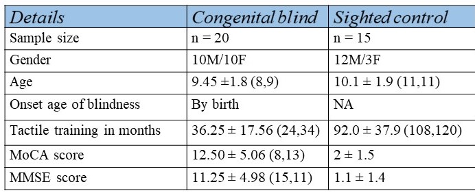
High-resolution T1W images and task based functional MR images of 20 congenital and 15 sighted control subjects (Table 1) were acquired using 3T MR scanner (Achieva 3T TX). Braille reading task comprised of 16 nouns (followed by four synonym options for each word), presented in block design, with Blood oxygenation level dependent (BOLD) signal measurement in 192 dynamics (TR/TE: 2000/30ms, 29 contiguous slices, 4.5mm thickness). The structural changes of cerebellum were evaluated using SUIT toolbox3 and Individual Component Analysis using CONN toolbox.4,5 Influence of language and cerebellum areas on task-based fMRI was investigated using three connectivity measures (global connectivity, local connectivity and cluster coefficient task). Correlation of functional connectivity measure were done with duration of Braille reading.Results
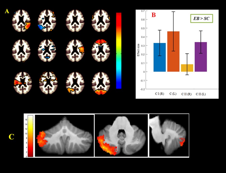
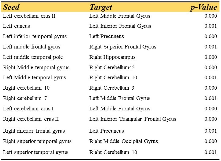
Seed to target analysis for ROI analysis exhibit increased FC between the posterior cerebellar (pCP), visual and language networks (LN). Differences in FC associated with duration of exposure of language semantic were observed in visual (VN), cerebellar (CP; CA), and language components (Table 2, Figure 1). Increased BOLD activation and gray matter volume were observed in cerebellar lobule in Vermis 3, lateral portion of Crus I and II and posterior cerebellar areas of CB compared to SC. A positive correlation of BOLD and FC measures with duration of Braille reading was observed in congenital blind.Discussion
Increased functional connectivity was observed between language regions (inferior frontal gyrus, superior temporal gyrus, middle temporal gyrus, inferior temporal cortices, precuneus) and bilateral cerebellar areas (crus I, crus II, cerebellum 45). Functional involvement of visual cortex, IFG, STG, MTG and cerebellum during braille reading task may be attributed to language processing.1,2 Functional connectivity of cerebellum with visual cortex, prefrontal, MTG and hippocampus in congenital blind group exhibit intact language perception and processing networks and increased correlation with duration of Braille reading may be attributed to non-visual processing involving comprehension of complex semantics and encoding of concrete words.1,6,7 Considering the tactile based Braille reading and enhanced connectivity involving the multi-sensory regions, results reflect a compensatory mechanism in congenital blind.Conclusion
Increased strength of connectivity and BOLD activation in cerebellum suggest intact language processing through fluency and decoding of haptic language.Acknowledgements
The authors acknowledge Department of Science and technology, Government of India for funding the study.References
1. Amedi A, Malach R, Hendler T, Peled S, Zohary E. Visuo-haptic object-related activation in the ventral visual pathway. Nat Neurosci. 2001;4(3):324–30.
2. Buchel C. Functional neuroimaging studies of Braille reading: cross-modal reorganization and its implications. Brain. 1998;121(7):1193–4.
3. Diedrichsen, J. et al. Imaging the Deep Cerebellar Nuclei: A Probabilistic Atlas and Normalization Procedure. NeuroImage. 2011;54(3): 1786–94.
4. Whitfield-Gabrieli S, Nieto-Castanon A. Conn : A Functional Connectivity Toolbox for Correlated and Anticorrelated Brain Networks. Brain Connect. 2012;2(3):125–41.
5. Friston KJ, Holmes AP, Price CJ, Büchel C, Worsley KJ. Multisubject fMRI studies and conjunction analyses. Neuroimage. 1999;10(4):385–96.
6. Stoodley, C. J., & Schmahmann, J. D. The cerebellum and language: evidence from patients with cerebellar degeneration. Brain and Language. 2009;10(3), 149–53.
7. Wallentin M, Ostergaard S, Lund TE, Ostergaard L, Roepstorff A: Concrete spatial language: see what I mean? Brain Lang 2005, 92: 221–233.
Figures