3010
Volumetric estimation of various brain parts in Gluten Ataxia patients: A quantitative MRI study1Nuclear Magnetic Resonance and MRI Facility, All India Institute of Medical Sciences, New Delhi, India, 2Pathology, All India Institute of Medical Sciences, New Delhi, India, 3Neurology, All India Institute of Medical Sciences, New Delhi, India, 4Gasteroenterology and Human Nutrition, All India Institute of Medical Sciences, New Delhi, India
Synopsis
Present study investigated volumetric volume changes in the brain of patients suffering from Gluten Ataxia (GA) using MRI. GA patients were seen to have significantly low brain and cerebellar volumes along the lobules which form the part of vermis suggesting cell degeneration and role of vermis in GA. GA patients also had significantly high levels of CSF. No significant changes were observed in whole brain grey matter, cerebrum, caudate, hippocampus and amygdala. Our results suggest that cerebrum volume is not linked to GA but lobes of cerebellum, whole brain white matter and CSF is significantly associated with GA.
Introduction
Gluten ataxia (GA) is a rare immune-mediated disorder that is caused due to immunological response towards ingested gluten in genetically susceptible individuals1,2. It accounts for upto 40% of the cases of idiopathic sporadiac ataxia and 15 % of all ataxias3. Symptoms of the disease are mild at onset but may lead to permanent neuronal damage, if not identified and managed on time. Actual neuropathology of the disease is not known but it presents with ataxia of gait and damage to the cerebellum. The potential role of the cerebellum in the neuropathology of gluten ataxia has been explored4. However, it is also suggested that some patients with GA have sensory (related to cerebrum) rather than cerebellar ataxia because of the absence of atrophy of the cerebellum on magnetic resonance imaging (MRI) in 40% of patients5. Role of the cerebrum is to initiate and co-ordinate movement and to control senses and any change in the volume of cerebrum can directly affect the senses and movement. So, there is a need to find out if there are any volume changes in the cerebrum of GA patients that is contributing to difficulty in movement. Volumetric information gathered from the Magnetic MRI data will be able to show different brain regions that are affected due to GA. Till date, no study has been done that has compared the volume of cerebrum in GA patients. Thus, present study aimed to investigate the changes in volume of various brain parts including cerebrum and cerebellum due to GA.Objective
To perform volumetric estimation of whole brain in the patients of GA using MRI and to compare the results with healthy controls to find if any brain part other than cerebellum is involved in GA.Patients and Methods
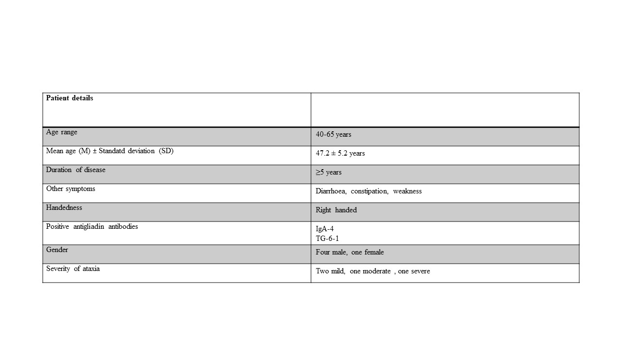
Patients The GA patient group (sporadic ataxia with positive antigliadin antibodies) consisted of 5 patients recruited from the fAtaxia Clinic, Department of Neurology of our Institute. Control group consisted of 10 healthy volunteers with no neurological/psychiatric disease/no medical or family history of Ataxia. The diagnosis of GA was made on the basis of clinical features, positive serological markers (antigliadin antibodies IgA and IgG, TG-2 and TG-6) and genetic testing of DNA. The study was approved by Institute Ethics Committee, and written informed consent was obtained from each subject. Details of neurological history and any background history of autoimmune disorders were recorded, duration of ataxia and requirement of mobility aids at the time of recruitment. Genetic testing for inherited spinocerebellar ataxias (SCA 1,2,3,6,7) and Friedreich’s ataxia (FA) were done and the patient was excluded if any of the inherited ataxias was found. Magnetic Resonance Imaging Volumetric 3-D T1 and T1-weighted images in all three planes, axial, coronal and sagittal of whole brain were acquired using standard spin-echo pulse sequence at 3T (Ingenia, Philips). The images were processed with the web based online tool volBrain pipeline and CERES that provides automated brain segmentation and generates report summarising the volumetric results. The percentage [(Mean±standard deviation (SD)) of different brain volumes were calculated and compared between the two groups using student t-test (SPSS, Chicago, Illionois). All volumetric analysis results are reported as a % of total intracranial volume. The level of significance was set at p<0.05.Results
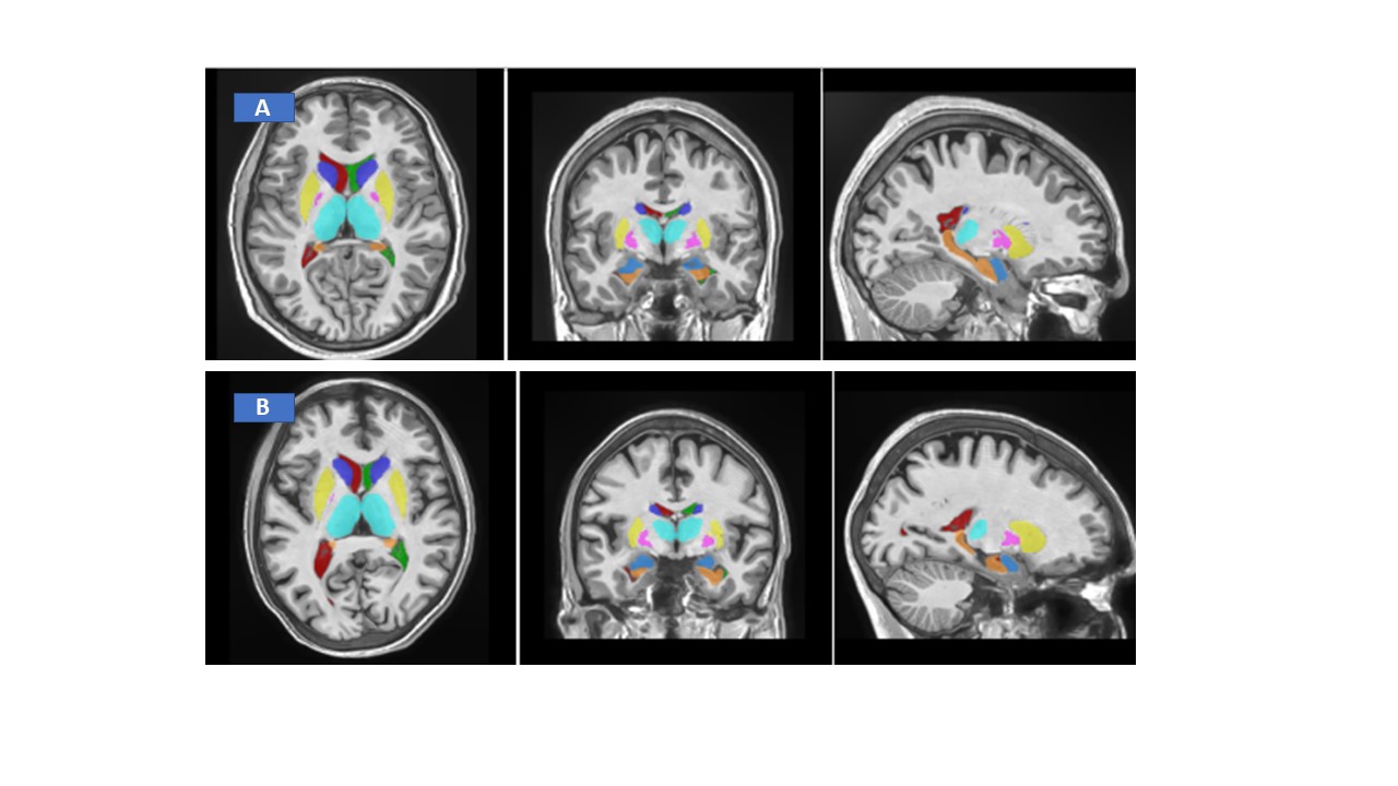
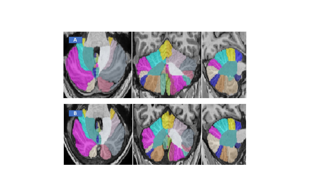
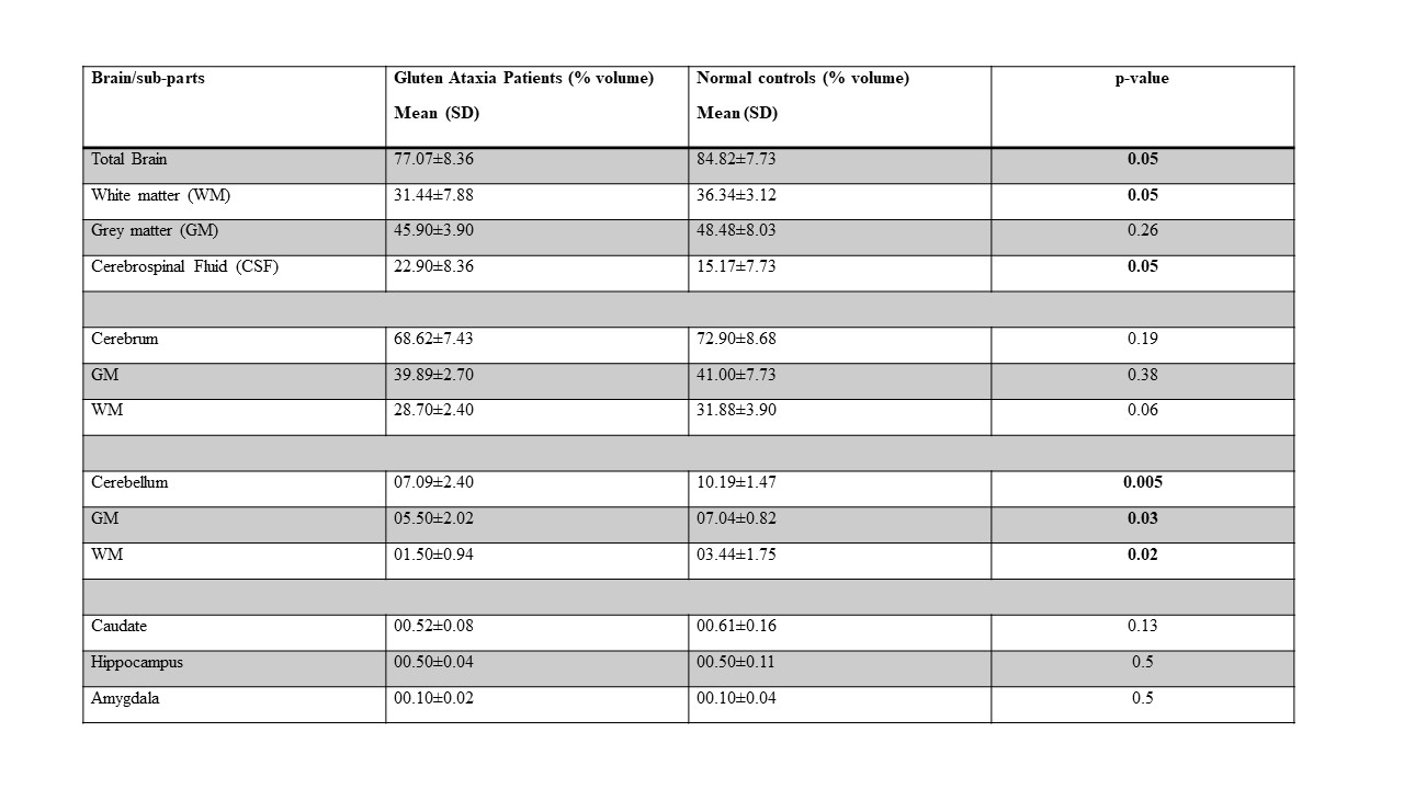
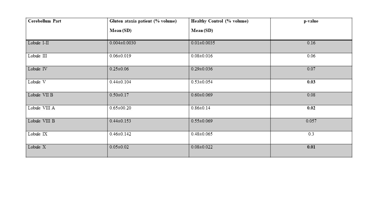
Figures 1 and 2 show the volumetric segmentation of whole brain and its parts. The results of volumetric analysis of the structure of the whole brain, its different parts, whole cerebellum and its components showed that compared to healthy controls, the whole brain volume was reduced in case of GA patients, including white Matter, cerebrospinal fluid, total Brain and cerebellum (Table 2). Cerebrum volume, volume of caudate, hippocampus and amygdala were not found to be different in the two groups. The total cerebellum volume and volume of cerebellum lobes V, VIII A and X was decreased in case of GA patients (Table 3).Discussion
Present study demonstrated significant changes in various brain parts in patients with GA compared to controls. The main findings of our study are significantly lower brain volume in GA patients including total brain volume, cerebrospinal fluid, cerebellum, grey and white matter, lobules V, VIII A and X of cerebellum. GA patients were seen to have significantly low brain and cerebellar volumes along the lobules which form the part of vermis suggesting cell degeneration and role of vermis in GA. GA patients also had significantly high levels of CSF volume indicating infection or injury. No significant changes were observed in cerebrum, caudate, hippocampus and amygdala. The clinical characteristics of gluten ataxia are not distinctive enough to allow clinical recognition from other ataxias and other neurodegenerative diseases. Our MRI findings, demonstrated atrophy of various brain parts in patients with GA that may have the clinical relevance by aiding diagnosis of these patients.Conclusion
Our results suggest that cerebrum volume is not affected in GA patients but lobes of cerebellum, whole brain white matter and CSF is significantly reduced in GA.Acknowledgements
The authors acknowledge intramural funding (A676) provided by All India Institute of Medical Sciences, New Delhi.References
1. Hadjivassiliou M, Davies-Jones GA, Sanders DS, Grünewald RA. Dietary treatment of gluten ataxia. J Neurol Neurosurg Psychiatry. 2003;74(9):1221-1224.
2. Hadjivassiliou M, Grünewald R, Sharrack B, et al. Gluten ataxia in perspective: epidemiology, genetic susceptibility and clinical characteristics. Brain. 2003;126(Pt 3):685-691. doi:10.1093/brain/awg050
3. Hadjivassiliou M, Grünewald RA, Chattopadhyay AK, et al. Clinical, radiological, neurophysiological, and neuropathological characteristics of gluten ataxia. Lancet. 1998;352(9140):1582-1585. doi:10.1016/s0140-6736(98)05342-2
4. Currie S, Hadjivassiliou M, Craven IJ, Wilkinson ID, Griffiths PD, Hoggard N. Magnetic resonance imaging biomarkers in patients with progressive ataxia: current status and future direction. Cerebellum. 2013;12(2):245-266. doi:10.1007/s12311-012-0405-3
5. Bürk K, Bösch S, Müller CA, et al. Sporadic cerebellar ataxia associated with gluten sensitivity. Brain. 2001;124(Pt 5):1013-1019. doi:10.1093/brain/124.5.1013
Figures