Shifeng Tian1, Ailian Liu1, Yan Guo2, and Yuan Wei1
1Department of Radiology, The First Affiliated Hospital of Dalian Medical University, Dalian, China, China, 2GE Healthcare, Dalian City, China, China
Synopsis
Squamous cell carcinoma is the most common pathological type in cervical cancer, followed by cervix adenocarcinoma. There are differences in the evaluation of prognosis between the two. Radiomics can quantitatively analyze a large number of image data, and then quantify tumor heterogeneity and noninvasively evaluate tumor biological behavior. The results suggest that radiomics based on mean kurtosis maps of diffusion kurtosis imaging sequences can identify different types of cervical cancer.
INTRODUCTION
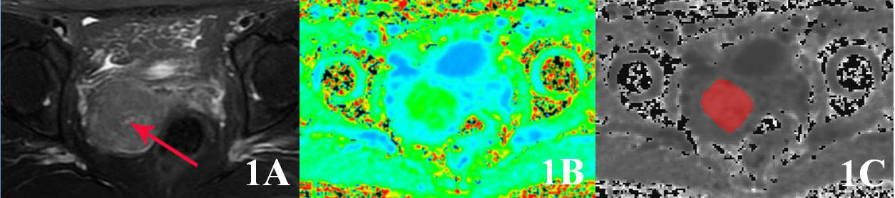
Compared with squamous cell carcinoma, cervix adenocarcinoma is often concealed, and early diagnosis is difficult. It is more likely to infiltrate the deep stroma and involve the vascular space, resulting in lymph node and distant metastasis. The survival rate of patients is lowand the sensitivity to radiotherapy is poor[1]. Therefore, it is valuable to accurately predict the pathological types of cervical cancer before operation. The diffusion kurtosis imaging can accurately reflect the changes of microenvironment in the tissue and quantify the deviation of water molecules due to non-Gaussian distribution and diffusion. The mean kurtosis is the representative parameter [2] of the DKI. Radiomics uses advanced image processing technology to extract high-throughput image feature information from imaging data, and analyze clinical information such as disease prediction and analysis through feature screening and model construction. The purpose of this study is to explore the application value of radiomics based on DKI sequence MK map in identifying CSCC and CA.METHODS
Retrospectively collected 63 patients with cervical cancer who underwent 1.5T MRI (SignaHDxt, GE Medical Systems, USA) examination (including DKI sequence) in our hospital from January 2017 to December 2019 and confirmed by surgical pathology. And there are 42 cases of CSCC and 21 cases of CA. Patients were divided into training group (n=43) and test group (n=20). Using ITK-SNAP software, ROI was manually delineated on MK image layer by layer, and then 3D-ROI of tumor was synthesized. Then it was imported into A.K. analysis software for feature extraction of high-dimensional imageology, and correlation test and gradient boosting decision tree were used for feature selection and dimension reduction. Then we built a multiple logical regression model and drawing ROC curves to evaluate the diagnostic effectiveness of the model and verify its effectiveness in the test group.RESULTS
A total of 386 imaging features were extracted, and 7 omics characteristics related to cervical cancer pathological classification were obtained by dimension reduction. The accuracy, AUC, sensitivity, and specificity of the constructed model for differentiating different pathological types of cervical cancer are 74.4%,0.867,85.7%,69.0% in the training group, and 80.0%,0.846,85.7%,76.9% in the test group.DISCUSSION AND CONCLUSIONS
Different radiomics features can indirectly reflect the heterogeneity of tumors.There are differences between CSCC and CA in cell arrangement, cell density and extracellular space, etc., which makes the image texture of the two different, sothere are differences in multiple omics features. Radiomics based on DKI sequence MK map can effectively identify different pathological types of cervical cancer and contribute to clinical decision-making.Acknowledgements
No acknowledgement.References
[1]Lavaud P, Fedida B, Canlorbe G, et al. Preoperative MR imaging for ESMO-ESGO-ESTRO classification of endometrial cancer. Diagn Interv Imaging, 2018, 99(6):387-396.
[2] Hempel JM, Brendle C, Bender B, et al. Diffusion kurtosis imaging histogram parameter metrics predicting survival in integrated molecular subtypes of diffuse glioma: An observational cohort study. Eur J Radiol, 2019, 112:144-152.