2996
Is it Feasible? IVIM-DWI and T2WI-based Texture Analysis Predicting Histological Types of Cervical Carcinoma Before Operation1The First Affiliated Hospital of USTC, Anhui Provincial Cancer Hospital, Hefei, China
Synopsis
The combination of IVIM-DWI biomarkers and T2WI-based texture features had good predictive performance to evaluate three different histological types of cervical carcinoma by synergizing diffusion, perfusion and heterogeneous features, especially for cervical squamous cell carcinoma and adenocarcinoma. A novel quantitative preoperative imaging model composed of IVIM-DWI and TA is a useful supplement for histopathological diagnosis, and might be used in clinical practice to assist in the formulation of treatment strategy.
INTRODUCTION
Cervical carcinoma, with the highest mortality rates among females in China, is one of the three most malignant tumor in female reproductive system1,2. Importantly, effective diagnosis of preoperative histological type may lead to better prognosis. With improved diagnosis on different histological types of cervical carcinoma, patients could receive adequate individualized treatment targeting different histological tumors 3. Although pathology is the golden standard of diagnosis on cervical carcinoma, the spatial heterogeneity occurred in different regions cannot be comprehensively obtained in routine cervical local biopsy 4. However, the heterogeneity of tumor might pose a pivotal challenge for histological diagnosis, treatment strategy and prognosis 5. Therefore, the techniques focusing on intratumor heterogeneity and microenvironment changes of cervical carcinoma are certainly worth more studies to improve the predictive accuracy of the whole lesion.Intravoxel incoherent motion diffusion-weighted imaging (IVIM-DWI), as a noninvasive quantitative molecular imaging technique, provides estimation of the diffusion motion of water molecules with higher b values (> 200 s/mm2) as well as the perfusion information of capillary microcirculation with lower b values (< 200 s/mm2) 6. The texture analysis (TA), as an advanced tool to reflect heterogeneity of tumors, it gathers the information of intensity, orientation, distribution and other features of the imaging voxels in lesions. MRI-based texture analysis may provide more quantitative information for predicting the heterogeneity of lesions 7.
In the present study, we aim to evaluate the predictive performance of IVIM-DWI biomarkers combined with T2WI-based texture analysis for different histological types of cervical carcinoma. A novel predictive paradigm was developed by synergizing the predictive capacity of diffusion, perfusion and heterogeneous features of cervical carcinoma.
METHODS
A total of 121 pathologically proved patients with cervical carcinoma (87 squamous cell carcinoma, Group-1/G-1; 28 adenocarcinoma, Group-2/G-2; 6 small cell carcinoma, Group-3/G-3) underwent IVIM-DWI to obtain ADC, D, D* and f. The whole lesion on T2WI was sketched manually to capture 18 histogram, 24 gray level co-occurrence matrix, 16 gray level size zone matrix and 16 gray level run length matrix features. All variables were analyzed by the steps of univariable and multivariable analyses.RESULTS
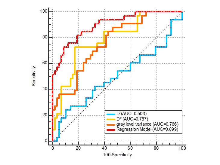
The univariable analysis revealed that the comparison of ADC, D*, f and 29 texture features were statistically significant between G-1 and G-2 (AUCG-1vsG-2, 0.654–0.795; p < 0.05). The comparison of ADC, D and 5 texture features reached significant levels between G-1 and G-3 (AUCG-1vsG-3, 0.623–0.956; p < 0.05). The ADC, D and 2 texture features of G-2 differed significantly compared to G-3 (AUCG-2vsG-3, 0.784–0.926; p < 0.05). In multivariable logistic regression analysis, the independent predictive factors for G-1 vs. G-2 were D, D* and gray level variance. The combination of IVIM-DWI and T2WI-based texture features improved the diagnostic value compared with individual parameters for the differentiation of cervical squamous cell carcinoma and cervical adenocarcinoma (AUC, 0.899; pairwise comparison p < 0.05).DISCUSSION
I.The Predictive Performance of IVIM-DWI.According to the results of our study, the ADC value composed of all diffusion coefficients related the resulting motion could differentiate three histological groups. It reflected the comprehensive effect of diffusion and perfusion information in tissues, and proved the differential diagnostic value of conventional DWI, just as the previous studies 8. Additionally, with the results of D value, we concluded that the pure water molecular diffusion motion, excluding the effect of blood flow microcirculation perfusion of different lesions, might be more accurate to reflect the diffusion information in tumor tissue for cervical small cell carcinoma comparing to cervical squamous carcinoma and adenocarcinoma. Previous studies also got the conclusion that the differences between the cervical squamous cell carcinoma and adenocarcinoma could be detected by IVIM-DWI 9.
According to the diagnostic efficacy of IVIM-DWI parameters for different histological groups, the D had a highest AUC of 0.956 for the comparison of G-1 and G-3, and the ADC had best performance for the comparison of G-2 and G-3 (AUC, 0.926). For the comparison of G-1 and G-2, the AUCs were 0.662 to 0.787, which could be improved by joint efforts of IVIM-DWI combined with T2WI-based texture features.
II.The Predictive Performance of TA and the Combination Value of IVIM-DWI and TA Techniques.
We tested 74 texture features on T2WI. The reason why we chose T2WI for TA was its high sensitivity to pathological processes 10. And from a preliminary experiment of our previous study, we found the texture features on T2WI had a higher diagnostic performance than DWI (b = 800 s/mm2) images.
For the comparison of G-1 and G-2, we found 29 texture features had differential diagnostic value (p < 0.05; AUC, 0.654–0.795). Then we formed a predictive model in multivariable logistic regression analysis composed of D, D* and gray level variance as independent predictive factors (AUC, 0.899). This model improved the predictive performance significantly by measuring the diffusion, perfusion and variance of gray level intensities together.
CONCLUSION
The combination of IVIM-DWI biomarkers and T2WI-based texture features had good predictive performance to evaluate three different histological types of cervical carcinoma by synergizing diffusion, perfusion and heterogeneous features, especially for cervical squamous cell carcinoma and adenocarcinoma. A novel quantitative preoperative imaging model composed of IVIM-DWI and TA is a useful supplement for histopathological diagnosis, and might be used in clinical practice to assist in the formulation of treatment strategy.Acknowledgements
This work was supported by the projects from the Foundation for Sci & Tech Research Project of Anhui Province, China (1804h08020294). We thank all staffs involved in the acquisition of data. We are grateful to all involved patients for their participation to the study.References
1. Manoharan D, Das CJ, Aggarwal A, et al. Diffusion weighted imaging in gynecological malignancies-present and future. World J Radiol 2016; 8(3): 288-298. https://doi.org/10.4329/wjr.v8.i3.288.
2. Tsikouras P, Zervoudis S, Manav B, et al. Cervical cancer: screening, diagnosis and staging. J BUON 2016; 21(2): 320-325.
3. Thapa D, Wang P, Wu G, et al. A histogram analysis of diffusion and perfusion features of cervical cancer based on intravoxel incoherent motion magnetic resonance imaging. Magn Reson Imaging 2019; 55: 103-111. https://doi.org/10.1016/j.mri.2018.06.016.
4. Gerlinger M, Rowan AJ, Horswell S, et al. Intratumor heterogeneity and branched evolution revealed by multiregion sequencing. N Engl J Med 2012; 366(10): 883-892. https://doi.org/10.1056/NEJMoa1113205.
5. Bedard PL, Hansen AR, Ratain MJ, et al. Tumour heterogeneity in the clinic. Nature 2013; 501(7467): 355-364. https://doi.org/10.1038/nature12627.
6. Wan Q, Deng YS, Zhou JX, et al. Intravoxel incoherent motion diffusion weighted MR imaging in assessing and characterizing solitary pulmonary lesions. Nat Publ Group 2017; 151: 1-8. https://doi.org/10.1038/srep43257.
7. Li z, Yu L, Wang X, et al. Diagnostic performance of mammographic texture analysis in the differential diagnosis of benign and malignant breast tumors. Clin Breast Cancer 2018; 18(4): e621-e627. https://doi.org/10.1016/j.clbc.2017.11.00.
8. Kuang F, Ren J, Zhong Q, et al. The value of apparent diffusion coefficient in the assessment of cervical cancer. Eur Radiol 2013; 23: 1050-1058. https://doi.org/10.1007/s00330-012-2681-1.
9. Becker AS, Perucho JA, Wurnig MC, et al. Assessment of Cervical Cancer with a Parameter-Free Intravoxel Incoherent Motion Imaging Algorithm. Korean J Radiol 2017; 18(3): 510-518. https://doi.org/10.3348/kjr.2017.18.3.510.
10. Dushyant VS, Anthony ES. Abdominal Imaging[M]. Saunders, Elsevier Inc 2011: 113.
Figures
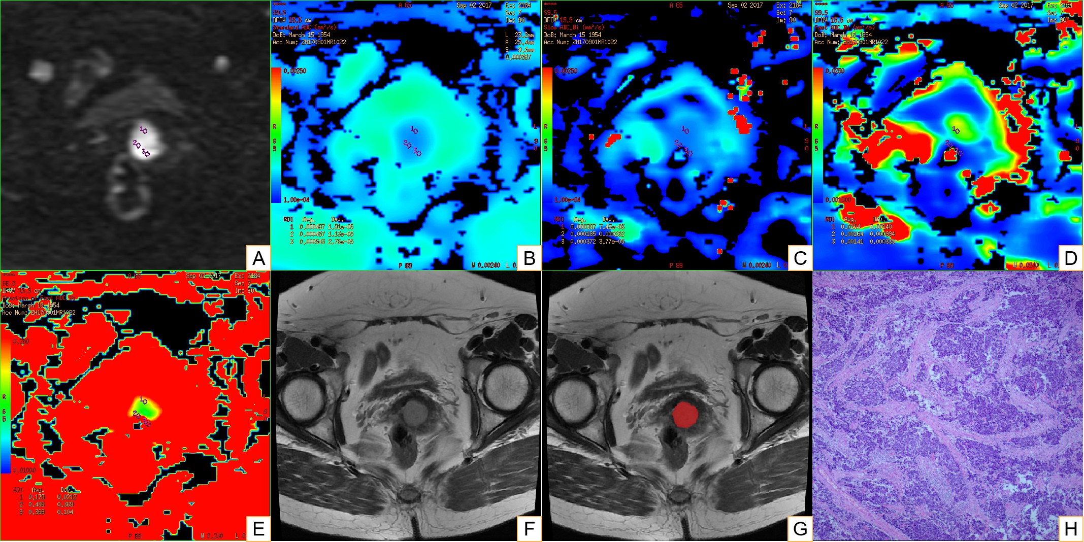
Fig. 1 Example of Manually Drawing ROIs for Cervical Lesion.
Panels A-H belong to a 63-year-old female with cervical small cell carcinoma. On panel A (IVIM-DWI at 1200 s/mm2), each radiologist drew ROI-1 (3 mm²) three times to get the values of ADC, D, D* and f, respectively (panels B-E). Panel F is the maximum area of the lesion on T2WI, and panel G was the manually drawing ROI-2 of the whole lesion. Panels H is the pathological performance, HE*400.
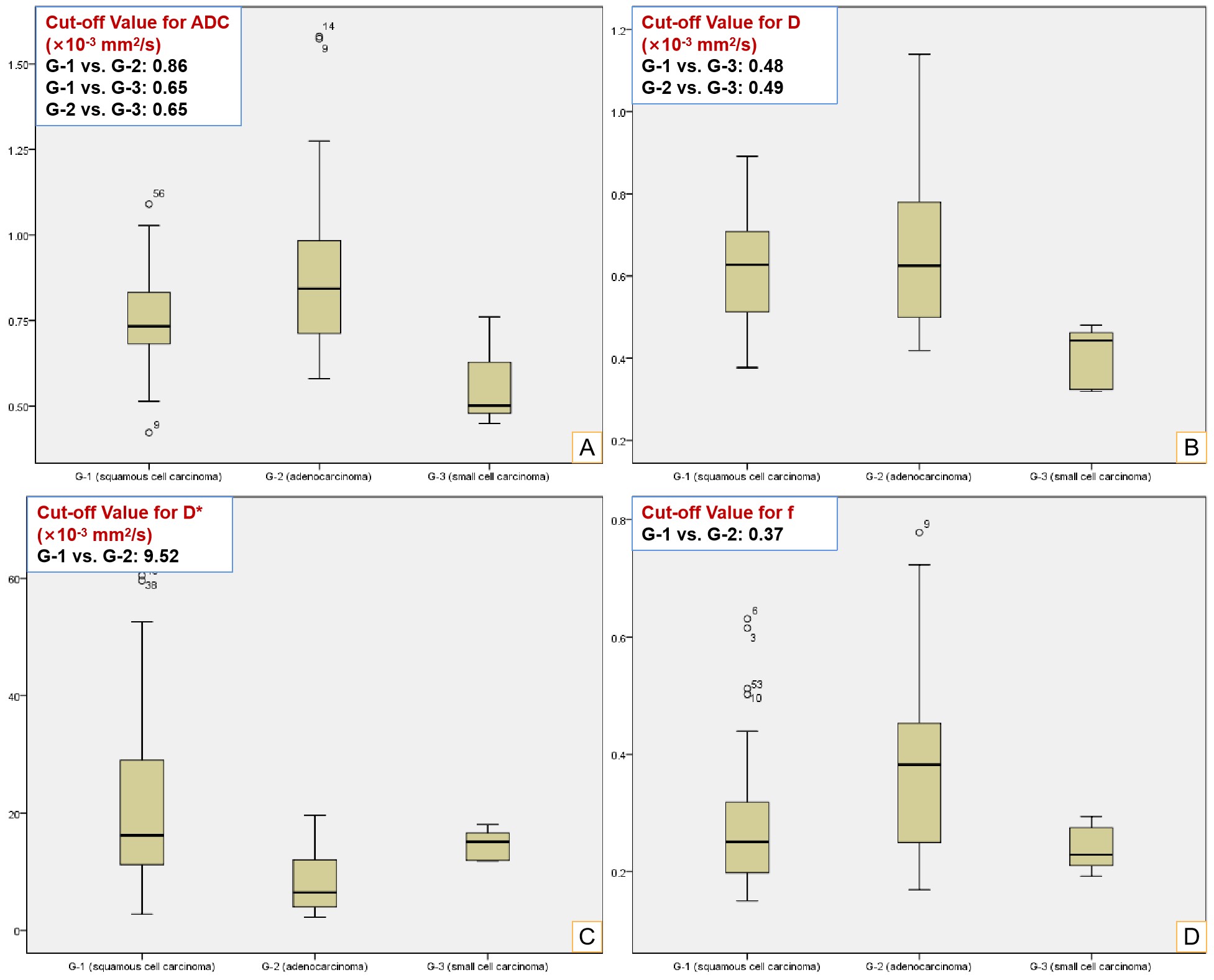
Fig. 2 Box-plots of IVIM-DWI Parameters.
Panels A-D represent ADC, D, D* and f values for G-1 (cervical squamous cell carcinoma), G-2 (cervical adenocarcinoma) and G-3 (cervical small cell carcinoma), respectively (p < 0.05).