2973
Ultra high resolution UTE imaging of the knee at 7T: Simultaneous view of cartilage, meniscus, ligament, tendon, and the chondro-osseous junction1Radiology, New York University, New York, NY, United States, 2Orthopaedic Surgery, New York University, New York, NY, United States
Synopsis
This human subject study presents preliminary results demonstrating the great potential of ultra-high resolution (0.14mm) UTE imaging to simultaneously visualize daily movement active tissues in the knee joint, including cartilages, menisci, ligaments, tendons, and even the chondro-osseous junctions. Non-invasive observation of these functional tissues are critical to understanding mechanisms of the onset and progression of osteoarthritis (OA).
INTRODUCTION
Recent studies have suggested that osteoarthritis (OA), especially post-traumatic OA, in the knee involves multiple functional components of the joint, such as cartilage, meniscus, ligament, tendon, and even the chondro-osseous junction.1-3 The onset and progression of OA may result from a comprehensive interaction among these functional tissues.4-5 It is unknown under what mechanisms these interactions lead to OA.6 Technical limitations hinder non-invasive studies on humans. Ultrashort echo time (UTE) MRI has the potential to non-invasively visualize these functional tissues of uniquely short T2 relaxation time.7-10 However, the UTE MRI performance is compromised at 3T when ultra-high resolution (0.14mm) is pursued to detect subtle defects and alterations in these tissues due to low signal-to-noise ratio (SNR).7 The 7T MRI doubles the SNR compared with the 3T 11 and would fully unleash the power of UTE imaging. Here we demonstrate such advantages for these critical functional tissues in the knee joint.METHODS
Five healthy subjects (age 28–58 years, male/female 2/3) were studied on a 7T whole-body MRI scanner (MAGNETOM 7T, Siemens) with a 28-channel Tx/Rx knee coil (QED, Ohio). IRB approval and written informed consents were obtained. A custom-developed 3D UTE sequence, acquisition-weighted stack of spirals (AWSOS),12 was used for data acquisition with optimized parameters: FOV=140x140x120 mm3, matrix size=1024x1024x60, resolution=0.14x0.14x2 mm3, flip angle=13°, fatsat=on, TE/TR=0.6/53ms, spirals =250, spiral readout Ts=16.72ms, and TA=13min34sec. The image reconstruction was implemented offline (due to the limited computation capacity of the scanner, version VB17A) with a custom-developed program in C++ (MS Visual Studio 2019, Redmond, WA). SNR was measured on magnitude images by taking a ratio of the mean intensity in a region of interest (ROI) to the scaled standard deviation (SD) in a noise-only background,13 i.e., SNR = MEAN*0.656/SD.RESULTS
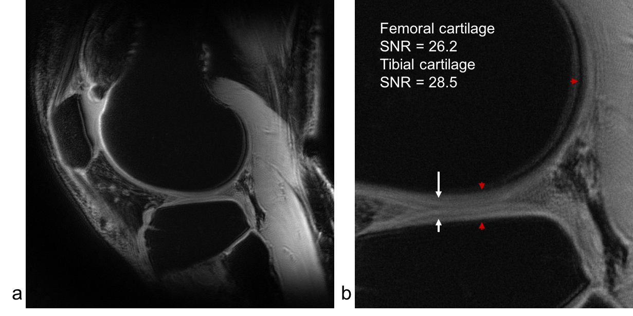
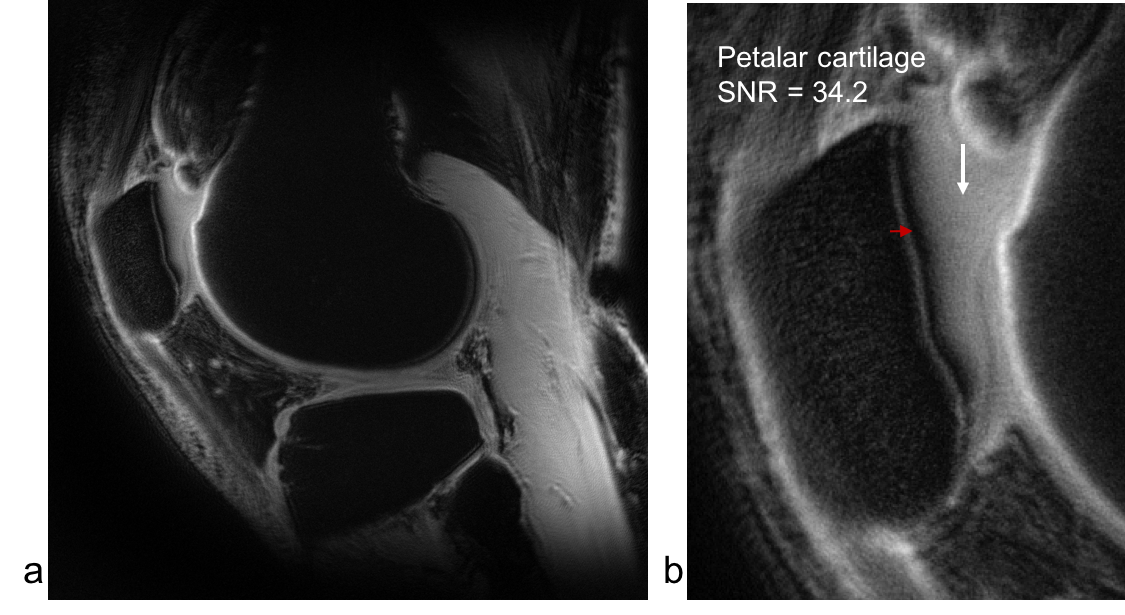
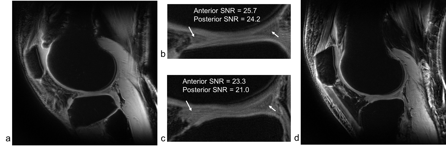
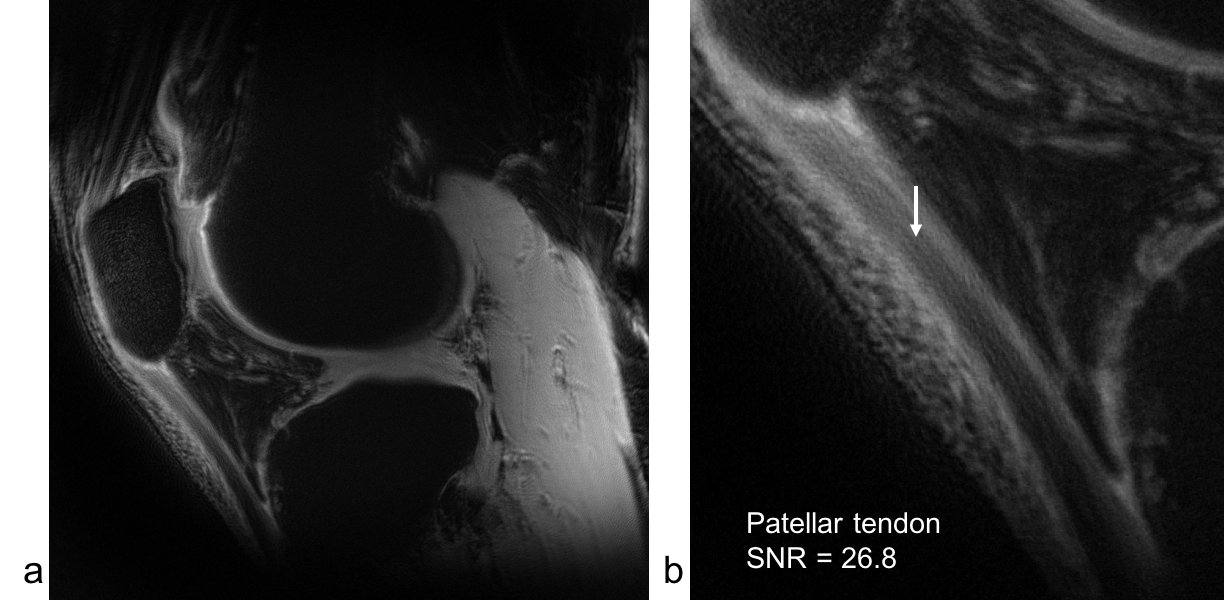
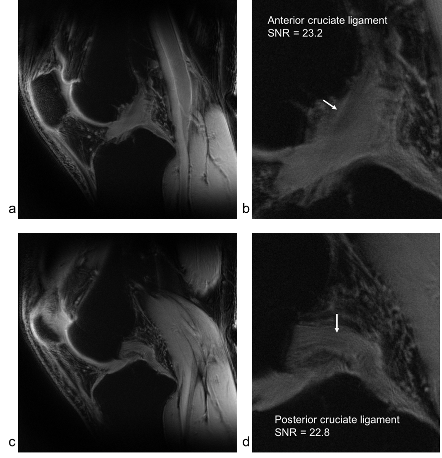
The ultra-high resolution UTE images at 7T for the studied subjects are shown in Figs. 1-5. They demonstrated the potential to simultaneously visualize multiple functional tissues in the knee joint: femoral and tibial cartilages (Fig. 1), patellar cartilage (Fig. 2), menisci (Fig.3), patellar tendon (Fig. 4), anterior and posterior cruciate ligaments (ACL and PCL, Fig. 5), and the chondro-osseous junction within the femur, tibia, and patella (Figs. 1, 2). The measured SNR of these tissues was between 22–34, which is high enough to visualize subtle alterations in morphology of these tissues. SNR in menisci across the male and female subjects is nearly unchanged (23.3 vs. 21.0), suggesting robustness and repeatability of the technique.DISCUSSION
Ultra-high resolution UTE imaging at 7T provided us with single scan for simultaneous visualization of multiple functional components of the knee joint in humans. This advantage mainly benefited from a matched combination of the AWSOS sequence, 7T magnet, and dedicated knee coil. Without them, the power of ultra high resolution UTE technique would not be fully revealed. The scan time (13min34sec) was acceptable as the studied subjects did not report any complaints, and were able to keep the knee still during the scan. However, SAR (specific absorption rate) was a major issue that led to a long TR (53ms) when fat saturation was on, compared with a shorter TR (20ms, and thus a shorter scan time 5min8sec) when fatsat was off.CONCLUSION
Ultra-high resolution UTE imaging at 7T has been shown to have the capability to visualize structural details of critical functional tissues in the knee joint of healthy subjects studied. It provides us with a new non-invasive imaging tool critical to understanding the mechanisms responsible for onset and progression of OA. More studies on subjects and OA patients are needed to consolidate this potential advantage.Acknowledgements
This work was financially supported by the General Research Fund of the Department of Radiology. This work was also performed under the rubric of the Center for Advanced Imaging Innovation and Research (CAI2R, www.cai2r.net), an NIBIB Biomedical Technology Resource Center (NIH P41 EB017183).References
1. Roemer FW, Kwoh CK, Hannon MJ, Hunter DJ, Eckstein F, Fujii T, Boudreau RM, Guermazi A. What comes first? Multitissue involvement leading to radiographic osteoarthritis, magnetic resonance imaging–based trajectory analysis over four years in the Osteoarthritis Initiative. ARTHRITIS & RHEUMATOLOGY 2015, 67 (8): 2085–2096. DOI 10.1002/art.39176.
2. Chu CR, Williams AA, West RV, Qian Y, Fu FH, Do BH, Bruno S. Quantitative Magnetic Resonance Imaging UTE-T2* Mapping of Cartilage and Meniscus Healing After Anatomic Anterior Cruciate Ligament Reconstruction. Am J Sports Med. 2014; 42(8):1847-1856.
3. Buckwalter JA, Brown TD. Joint injury, repair, and remodeling: roles in post-traumatic osteoarthritis. Clin Orthop Relat Res 2004; 423:7-16.
4. Lohmander LS. Articular cartilage and osteoarthritis. The role of molecular markers to monitor breakdown, repair and disease. J Anat 1994; 184: 477-492.
5. Chu CR, Williams AA, Coyle CH, Bowers ME. Early diagnosis to enable early treatment of pre-osteoarthritis. Arthritis Res Ther 2012;14(3):212.
6. Nelson AE, Allen KD, Golightly YM, Goode AP, Jordan JM. A systematic review of recommendations and guidelines for the management of osteoarthritis: The Chronic Osteoarthritis Management Initiative of the U.S. Bone and Joint Initiative. Seminars in Arthritis and Rheumatism 2014; 43:701–712.
7. Qian Y, Williams AA, Chu CR, Boada FE. High-resolution ultrashort echo time (UTE) imaging on human knee with AWSOS sequence at 3.0T. J Magn Reson Imaging 2012; 35(1):204-210.
8. Garwood MG, Idiyatullin DS, Corum CA, Moeller S. Frequency swept excitation for magnetic resonance. US Patent 8,067,936. Nov. 29, 2011.
9. Bydder M, Bydder GM, Robson M, Gatehouse P. Magnetic resonance imaging with ultra short echo times. US Patent 7,474,097. Jan. 6, 2009.
10. Pauly JM, Nishimura DG, Magnetic resonance imaging of short T2 species. US patent 5025216, 1991.
11. Springer E, Bohndorf K, Juras V, Szomolanyi P, Zbýň Š, Schreiner MM, Schmitt B, Trattnig S. Comparison of routine knee magnetic resonance imaging at 3 T and 7 T. Invest Radiol. 2017; 52(1):42-54.
12. Qian Y, Boada FE. Acquisition-weighted stack of spirals for fast high-resolution 3D UTE MR imaging. Magn Reson Med 2008; 60:135-145.
13. Kellman P, McVeigh ER. Image reconstruction in SNR unit: a general method for SNR measurement. Magnetic Resonance in Medicine 2005; 54:1439-1447.
Figures