2965
Deep, intermediate and superficial layers of patella cartilage assessment using T2 mapping in patients with chondromalacia.1Clinical and Research Institute of Emergency Pediatric Surgery and Trauma, Moscow, Russian Federation, 2National Research Nuclear University MEPhI, Moscow, Russian Federation, 3Philips Healthcare, Moscow, Russian Federation, 4Emanuel Institute of Biochemical Physics of RAS, Moscow, Russian Federation
Synopsis
In this work, the values of the transverse relaxation times (T2) were measured for the first time in the deep, intermediate, and superficial layers of the patellar cartilage at different stages of chondromalacia as compared to control. The T2 values in the intermediate layer of cartilage show the best sensitivity to the grade of injury and have statistically significant differences for the groups of normal, mild, and severe cartilage damage. The T2-mapping technique is a potential tool for studying structural changes in cartilage and T2 values can be considered as a non-invasive biomarker for the assessment of the cartilage condition.
Introduction
Chondromalacia patella is a pathological process that is associated with a thinning, softening, and gradual destruction of the cartilage on the posterior surface of the patella. This disease leads to a violation of the biomechanics of the patella and femur, which causes severe pain during movement. The main risk group is young people. Ineffective treatment of chondromalacia can lead to the early development of patellofemoral arthrosis and significantly impair the quality of life. Currently, the diagnosis of chondromalacia is based on a visual assessment of MRI images. There are some evidences that this method may underestimate the degree of cartilage damage. New non-invasive quantitative methods have been actively developing over the past few years. They are focused on understanding the biochemical composition of cartilage tissue. The largest part of the following works devoted to evaluation of cartilage tissue destruction in osteoarthritis using T2 mapping. The main aim of this work was studying of cartilage changes in patients with chondromalacia after patellar dislocation (common injury in youth sports). For this purposes T2 values were determined in the deep, intermediate, and superficial layers of the patellar cartilage depending on the grade of chondromalacia.Methods
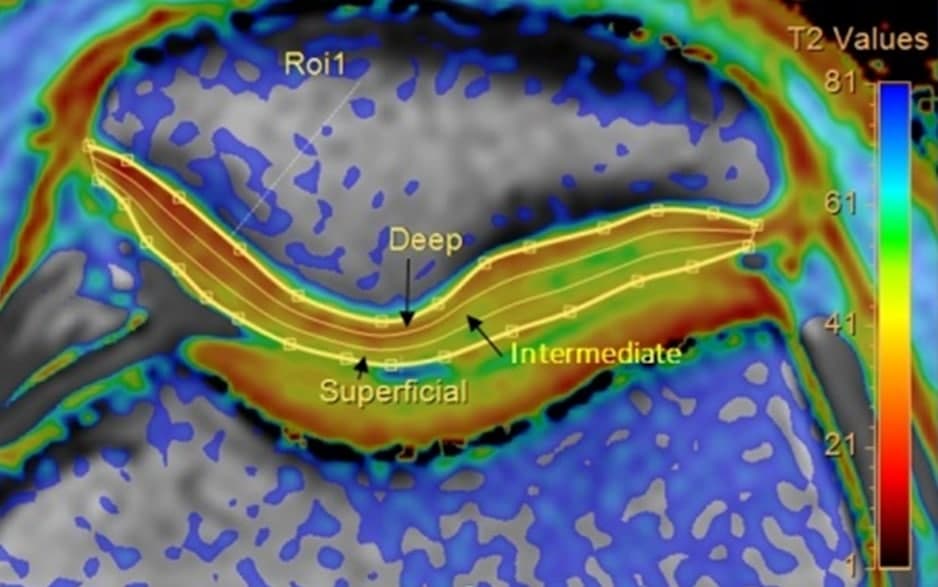
The study comprises 81 children (range: 12-17 years). The group of patients includes children hospitalized with acute lateral patella dislocation in Clinical and Research Institute of Emergency Pediatric Surgery and Trauma. All patients underwent MRI no later than 2-3 days after hospital admission. The control group consists of 14 age-matched healthy volunteers. MRI examination was performed at Philips Achieva dStream 3.0T scanner (Philips Healthcare) using 16-chanel dStream knee coil. The diagnostic protocol included routine MRI images, which were used by the radiologist to determine the grade of chondromalacia according to the modified Outerbridge scale. For further analysis, patients with the 1st and 2nd degrees of chondromalacia were grouped into the mild trauma group (total - 52 patients) and patients with the 3rd and 4th degrees - into the severe trauma group (total - 15 patients). The scientific protocol included T2 mapping (TSE sequence, 13 TE in the range 9-72 ms, TR = 2000 ms, NSA = 2). The Cartilage Assessment utility in the Philips IntelliSpace Portal software was used for the quantitative T2-maps analysis. The T2 values were obtained in each of the three evenly separated cartilage layers (Fig. 1). The Mann-Whitney U-test was used to reveal intergroup differences between patient groups. The threshold of significance was set at p<0.05.Results
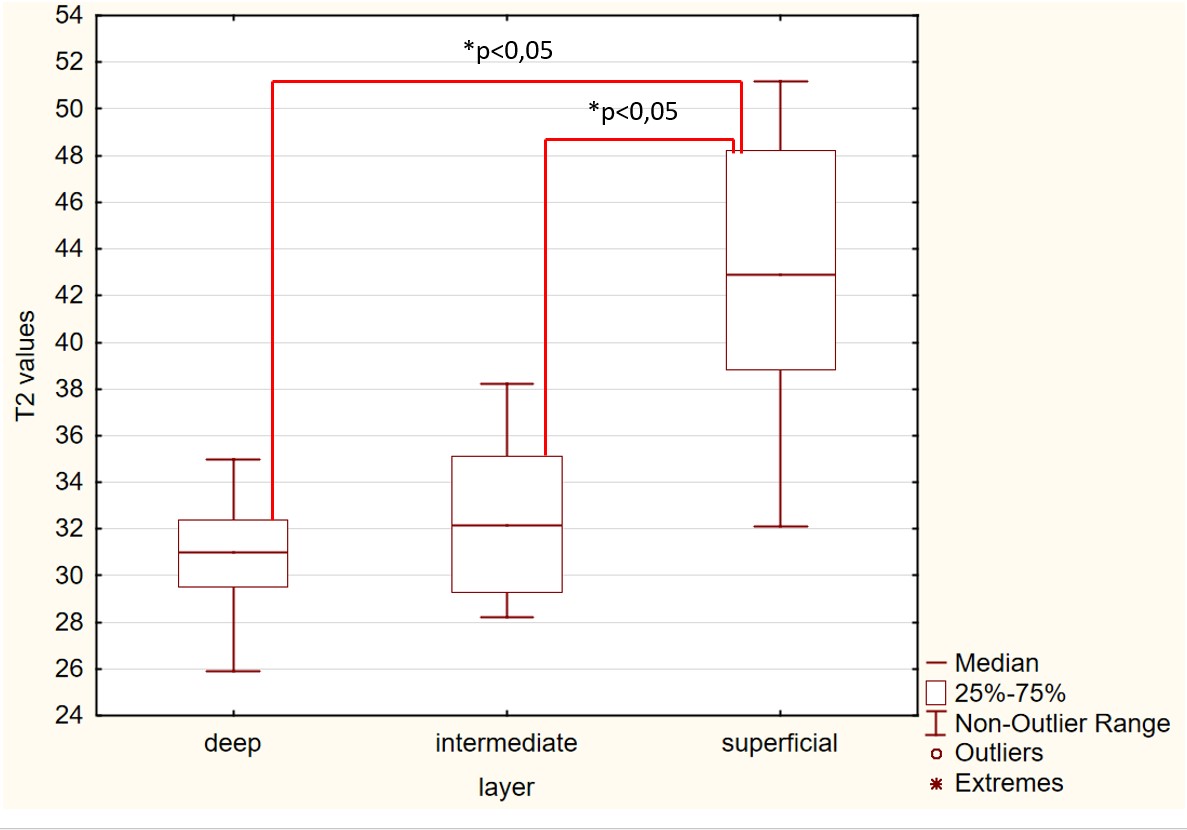
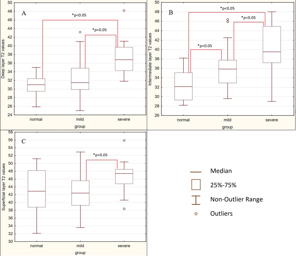
The results for the normal group are presented in Fig. 2. It has been found that the T2 values in the surface layer are significantly higher than in the other two layers. The comparison results of the T2 values in patients of the normal group, the mild injury group, and the severe injury group are shown in Fig. 3. There is statistically significant T2 values increase in the deep and intermediate layers in patients with severe trauma compared to the normal group. In the intermediate layer, the transverse relaxation times are statistically distinguishable for the normal, mild injury, and severe injury groups – the more severe a damage, the higher T2 values.Discussion and conclusion
For the first time T2 values were determined in the deep, intermediate, and superficial patellar cartilage layers in patients with different stages of chondromalacia as compared with control group. An interest in measuring T2 relaxation times is based on their correlation with the biochemical composition of articular cartilage. According to the previous study1, T2 values are sensitive to water protons anisotropy in the cartilage tissue and reflect its concentration. The T2 sensitivity to collagen matrix integrity and collagen fiber orientation was also noted. The direction of the collagen fibers changes depending on the cartilage zone: they are parallel to the articular surface in the superficial zone, perpendicular in the deep zone, and have a random arrangement in the middle. The study of the normal group showed that T2 in the surface layer is significantly higher than in the deep and intermediate layers. It is not hard to guess that cartilaginous changes due to chondromalacia in different layers might be different. Thus, the layer-by-layer analysis of T2 values was carried out in our work. The results obtained for various degrees of chondromalacia showed the best sensitivity of T2 values to damage of the patellar cartilage in the intermediate layer – in our study transverse relaxation times are statistically distinguishable for normal, mild, and severe injury groups. The stronger the severity of the injury, the higher the T2 value. In the deep layer, T2 increases only in severe stages of chondromalacia, which is consistent with the results of study2. The increase in transverse relaxation times may result from the disorganization of collagen fibers, an increase in water content, and its mobility. Thus, it is essential to diagnostic purposes to quantify T2 values not from the entire volume of cartilage, but taking into account its structure, at least three layers – deep, intermediate and superficial layers. In conclusion, it should be noted that the T2-mapping method has potential for use in the evaluation of chondromalacia in children, as well as in evaluation of treatment effectiveness.Acknowledgements
No acknowledgement found.References
1. Mosher, T. J., Dardzinski, B. J. Cartilage MRI T2 Relaxation Time Mapping: Overview and Applications. Seminars in Musculoskeletal Radiology. 2004; 08(04): 355–368.
2. Väätäinen, U., Kiviranta, I., Jaroma, et al. Collagen Crosslinks in Chondromalacia of the Patella. International Journal of Sports Medicine. 1998; 19(02), 144–148.
Figures