2955
Morphologic Structure of Rabbit Suprapatella Cartilage by µMRI and PLM
Hannah Mantebea1, Syeda Batool1, Mouhamad Hammami1, and Yang Xia1
1Physics, Oakland University, Rochester, MI, United States
1Physics, Oakland University, Rochester, MI, United States
Synopsis
This study aims to quantity the structural morphology of sesamoid fibrocartilage in rabbits, using µMRI at 19.5-µm resolution and polarized light microscopy at 1-µm resolution. We show that the sesamoid fibrocartilage has one thin surface layer (~ 10 µm) of parallel-arranged fibers followed immediately by the majority of random fibers (~ 390 µm). T2 anisotropy was not observed in fibrocartilage. The collagen fibers in the surface layer are dense and in parallel with the surface, which allows it to withstand stress from the quadriceps tendon during flexion, while at the same time to reduce pressure load from the femur.
Introduction
Knee is a complex joint that consists of tibiofemoral and patellofemoral regions. In arthritis research using the rabbit model, the suprapatella in the knee, an essential weight bearing structure, was often overlooked despite its prominence and contribution (1). This study aims to quantity the structural morphology of sesamoid fibrocartilage in rabbits, using µMRI and polarized light microscopy (PLM).Methods
Sesamoid fibrocartilage from suprapatella (n=4) of 12-14 weeks-old New Zealand rabbits were studied quantitatively. µMRI experiments used a 7Tesla/89mm Bruker Avance IIIHD 300 NMR spectrometer. A quantitative T2 magnetization-prepared imaging sequence was used (2), using the echo times of 2, 4, 8, 10, and 12 ms, and with a 19.5-µm transverse resolution. Histological sections were made out of the µMRI-imaged specimens. The PLM system was a digital CCD camera mounted on a polarized light microscope (Leica, Germany). The output of the camera generates two quantitative images, the optical retardation and the angular orientation (3). Quantitative PLM images were obtained from thin sections at 1-µm pixel resolution.Results
Fig 1a shows a photo of the intact posterior aspect of suprapatella that articulates with the anterior aspect of the femur, and Fig 1b and 1c show the central-widthwise cut surface of suprapatella, a photo (1b) and viewed under a microscope (1c). Fig 2 shows three MRI T2-weighted intensity images of a suprapatella specimen at 19.5-µm resolution, at 0˚ and 55˚ to the magnetic field (vertically up). Quantitative T2 maps of the suprapatella (the rightest image) had a dark thin line on the surface region, which is likely due to the magnetic susceptibility difference between tissue and air since the tissue surface bounded by air in imaging. Fig 3a shows the quantitative 1D T2 profiles from these 2D images, which lacked the magic angle effect in the tissue (i.e., the 0˚ and 55˚ profiles were similar). Based on the T2 characteristics, the suprapatella cartilage seems to have a thin surface layer that covers the much thicker main tissue of about 390 µm in average thickness. Both layers in the suprapatella show little variation when the tissue blocks changed their orientations in the magnetic field. Fig 3b shows one set of quantitative PLM images at 1.0-µm pixel resolution. Several observations can be made from these quantitative results. First, the top 10 µm of the sesamoid fibrocartilage can be viewed as the surface layer (or skin) of the sesamoid fibrocartilage, which confirms the observation in µMRI. This skin has considerable portion of the collagen fibers being oriented in parallel with the surface of sesamoid fibrocartilage (as in the line drawings on the furthest right of Fig 3b), which would help in the tangential gliding when the joint is in motion. Below this organized skin, the quantitative values in most of the fibrocartilage have large variations, in contrast to the consistent zonal values in the PLM images for articular cartilage (3). The retardation values for most of the non-surface fibrocartilage varied around 1 nm, indicating the lack of a coherent fibril organizational structure.Discussions
In contrast to the triple structural zones in articular cartilage, the suprapatella only has two structural layers; a thin surface layer (~ 10 µm thick) and a much thicker main tissue. The collagen fibers in the surface layer are well organized, dense, and in parallel with the surface, which gives the surface layer with its short T2 values (Fig 3a). This suprapatellar surface layer likely aids to the reduction of friction when the surface slides with the femur cartilage. The majority of the suprapatella is its main tissue body, where the T2 values are consistently high and have no orientational dependence in the magnetic field. The high-resolution features of the quantitative µMRI and PLM results suggest the suprapatella has mostly the loosely packed fibers, together with mostly free water with insignificant dipolar interaction, making it more isotropic at the magic angle with respect to the magnetic field.Conclusion
The random arrangement of the fibers in the tissue offers flexibility to the suprapatella cartilage and allows it to withstand stress from the quadriceps tendon during flexion, while at the same time to reduce pressure load from the femur. To the best of our knowledge, this was the first quantitative imaging study that characterized the morphological structures of sesamoid fibrocartilage in the suprapatella of the rabbit at high resolutions. These quantitative results will be beneficial to future studies of joint diseases using rabbits as the animal model.Acknowledgements
Yang Xia is grateful to the National Institutes of Health (NIH) for a R01 grant (AR 069047). The authors are in debt to Mr. Farid Badar for the technical help in imaging, to Dr. Adam Lauver and Ms. Barbara Christian (Department of Pharmacology & Toxicology, Michigan State University, East Lansing, Michigan) for providing the rabbit samples.References
(1) T. Tischer, S. Milz, M. Maier, M. Schieker, and M. B. An immunohistochemical study of the rabbit suprapatella, a sesamoid fibrocartilage in the quadriceps tendon containing aggrecan. J. Histochem. Cytochem. 50, 955–960 (2002). (2) Xia, Y. (1998). "Relaxation Anisotropy in Cartilage by NMR Microscopy (µMRI) at 14 µm Resolution." Magn Reson Med 39(6): 941-949. (3) Xia, Y., et al. (2001). "Quantitative In Situ Correlation Between Microscopic MRI and Polarized Light Microscopy Studies of Articular Cartilage." Osteoarthritis Cartilage 9(5): 393-406.Figures
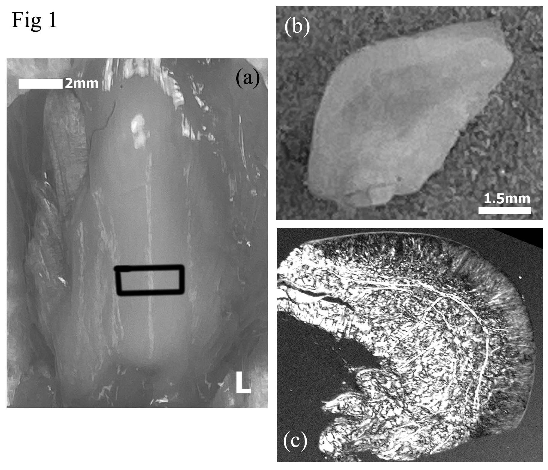
Fig 1 Photos and optical image
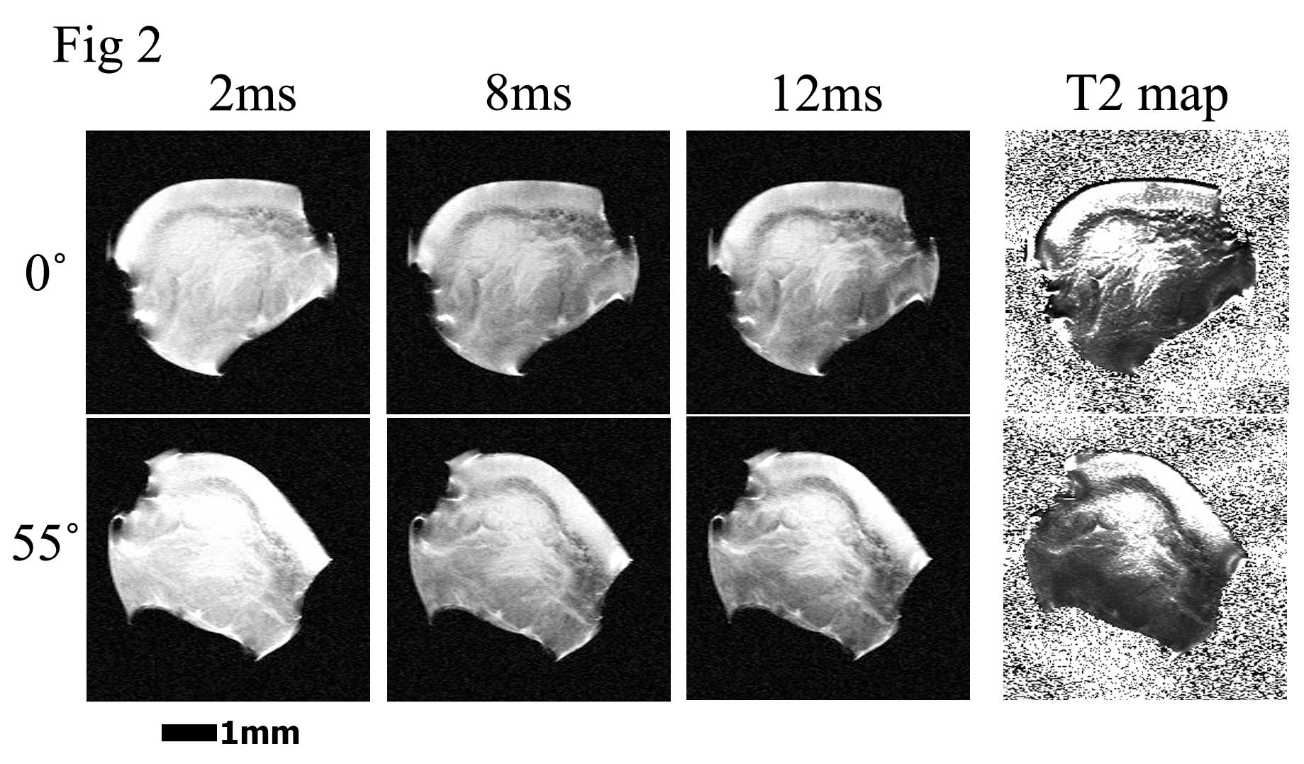
Fig 2 µMRI images
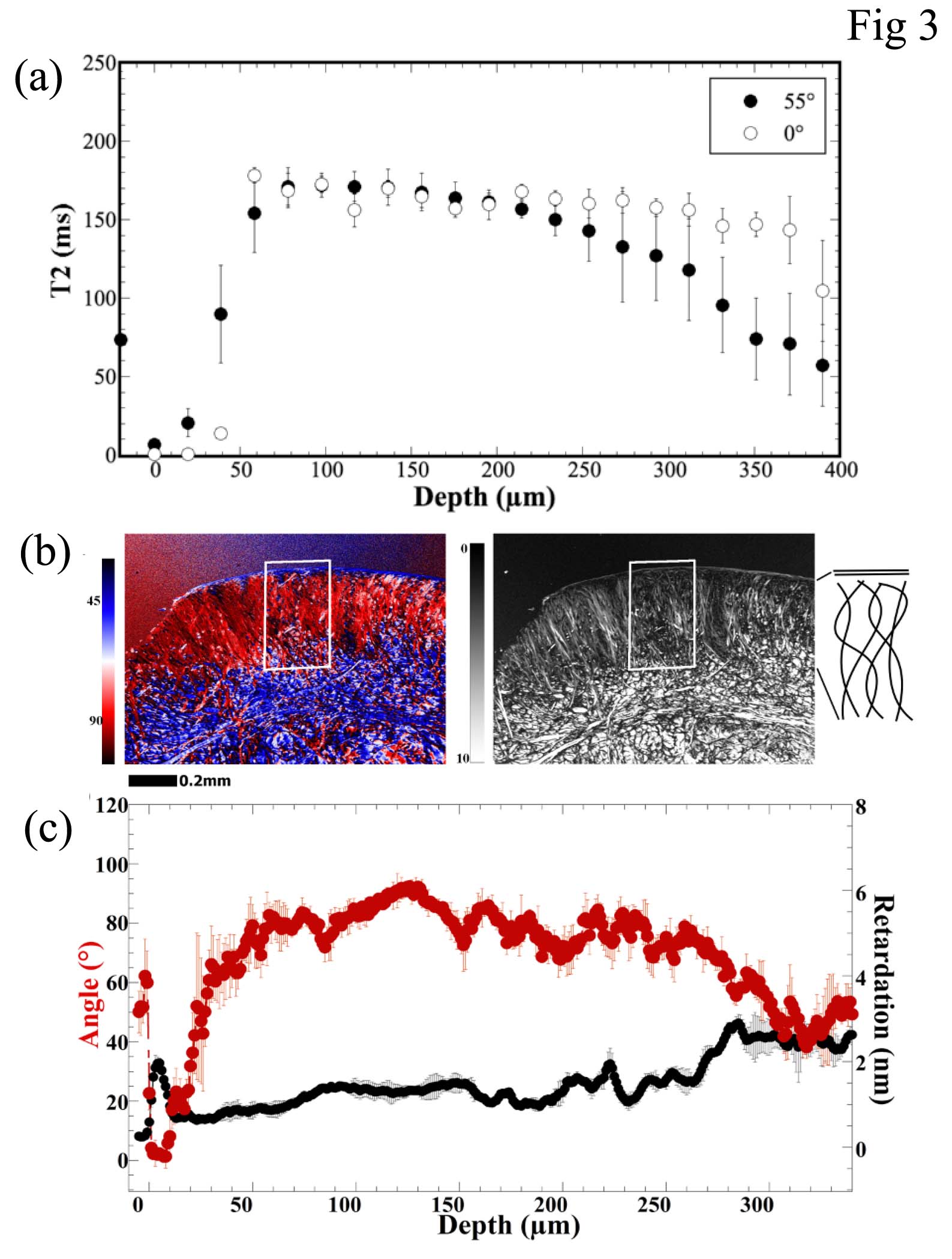
Fig 3 Quantitative µMRI profiles and PLM images/profiles