2934
Awake fMRI shows an impact of anesthesia on resting state functional connectivity in mice1Human Informatics and Interaction Research Institute, National Institute of Advanced Industrial Science and Technology (AIST), Tsukuba, Japan
Synopsis
This study aims to develop awake fMRI protocol to compare the resting state functional connectivity between awake state and anesthetized state (isoflurane or medetomidine + isoflurane) in the same mouse. The typical structure of resting state functional connectivity did not alter under ISO or MED+ISO anesthesia, but the connectivity strength and fractional amplitude of low frequency fluctuation in the cerebral cortex and the thalamic nuclei altered under anesthesia. These results indicate the availability of anesthesia on the resting state fMRI although the impact of anesthesia on neuronal activity should be considered.
Introduction
Anesthetic influences the neurovascular coupling as well as the neuronal activity. Although a few studies compared awake and anesthetized state in rodent fMRI1, the impact of anesthesia on the resting state functional connectivity has not been fully understood. In the current study, I developed awake fMRI protocol to compare the resting state functional connectivity between awake state and anesthetized state (isoflurane or medetomidine + isoflurane) in the same mouse.Methods
AnimalsMale C57BL/6 mice (20-30 g) (n = 18) were used.
FMRI experiment
FMRI acquisition was conducted at Bruker 11.7T with a cryoprobe. fMRI images were acquired using a gradient-echo EPI sequence with following parameters, TR/TE = 1,500/15 ms, spatial resolution = 150 x 150 x 500 µm3 / voxel, 15 slices, for 10 min (400 volumes). The schematic protocol of experiment is shown in Fig. 1. To perform awake fMRI, cranioplastic surgery was performed2. The cranioplastic acrylic cement was mounted on the skull with a plastic bar, which connected to a custom-made head positioner to fix the mouse’s head during the MRI session without pain. The mice were then performed on the acclimation training after 1 week from the surgery to adapt circumstance of MRI scanning. fMRI experiment started after acclimation training (4-7 days). The resting state fMRI data were acquired with awaked mouse following the adjustment of magnetic homogeneity and acquisition of anatomical image. Once fMRI acquisition was finished in awake state, mice were then anesthetized with 1.0% isoflurane (ISO) or 0.05 mg/kg/h medetomidine (i.p.) + 0.5% isoflurane (MED+ISO) in MR bore. Following 10-30 min after start of anesthesia, resting state fMRI data were acquired under anesthetized state. The respiration and the body temperature were maintained at the rate of 80 /min and 37 °C during the experiment. Anatomical images were acquired for spatial correction using multi-slice rapid acquisition with relaxation enhancement (RARE) following parameters: TE/effective TE = 2,500/48 ms, same FOV with high resolution (100 x 100 x 500 µm3 / pixel) and RARE factor = 8.
Image processing
The slice timing correction, spatial realignment, normalization and smoothing were performed by SPM12 (Welcome Trust Center for Neuroimaging, UK). Averaged signals in the CSF, the white matter and the head motion parameters were used as nuisance regressors. Detrending and temporal filtering (0.01 – 0.1 Hz) were then performed using CONN toolbox. For correlation analysis, 142 ROIs corresponding to Allen Brain Atlas was created. Time-course within each ROI was extracted and Pearson correlation coefficient between two ROIs was calculated. The fractional amplitude of low frequency fluctuation (fALFF) was calculated as the ratio of power within the frequency range between 0.01 and 0.1 Hz and of power over the total frequency range in each voxel in each mouse3.
Results
The overall averaged correlation coefficients under ISO or MED+ISO anesthesia was smaller than those under awake state (Fig. 2). Importantly, typical structure of the functional connectivity under anesthesia, such as interhemispheric connectivity and local connectivity within the cortex and thalamus, was observed both in anesthetized and in awaked state. The statistical analysis shows that correlation coefficients under ISO (Fig. 3A) and MED+ISO (Fig. 3B) significantly decreased rather than awake state, while there was no increase of correlation coefficient in anesthetized state. The fALFF was also investigated in anesthetized state and awake state. The fALFF in bilateral auditory cortex, somatosensory cortex, motor cortex and anterior cingulate cortex, hypothalamus increased and fALFF in the thalamic nuclei decreased under ISO anesthesia compared with the fALFF during awake state (Fig. 4A). The fALFF in bilateral auditory cortex, motor cortex, anterior cingulate cortex, entorhinal cortex and the hypothalamus increased rather than the fALFF during awaked state and fALFF in the thalamic nuclei decreased under MED+ISO anesthesia compared with the fALFF in awake state (Fig. 4B).Discussion
The present study shows an impact of light anesthesia on the resting state functional connectivity as well as the fALFF. The previous studies compared the functional connectivity under anesthesia using different animal group. In contrast, we compared the awake state and anesthetized state in the same animal and at the same experimental day. This protocol could directly show the effect of anesthesia in each animal. This result show that typical structure of resting state functional connectivity was observed under ISO and MED+ISO anesthesia, indicating the utility of light anesthesia for resting state functional connectivity. However, because entire connectivity strength and fALFF in the thalamus decreased both under ISO and under MED+ISO, the effect of light anesthesia on the neuronal activity should be considered in the resting state fMRI.Acknowledgements
No acknowledgement found.References
1. Stenroos P, Paasonen P, Salo RA, Jokivarsi K, Shatillo A, Tanila H and Gröhn O, Awake Rat Brain Functional Magnetic Resonance Imaging Using Standard Radio Frequency Coils and a 3D Printed Restraint Kit, Front. Neurosci., 20 August 2018.
2. Tsurugizawa T, Tamada K, Ono N, Karakawa S, Kodama Y, Debacker C, Hata J, Okano H, Kitamura A, Zalesky A, Takumi T, Awake functional MRI detects neural circuit dysfunction in a mouse model of autism, Sci Adv. 2020 Feb 5;6(6):eaav4520.
3. Nakamura Y, Nakamura Y, Pelosi A, Djemai B, Debacker C, Hervé D, Girault JA, Tsurugizawa T, fMRI detects bilateral brain network activation following unilateral chemogenetic activation of direct striatal projection neurons, NeuroImage. 2020 Oct 15;220:117079
Figures
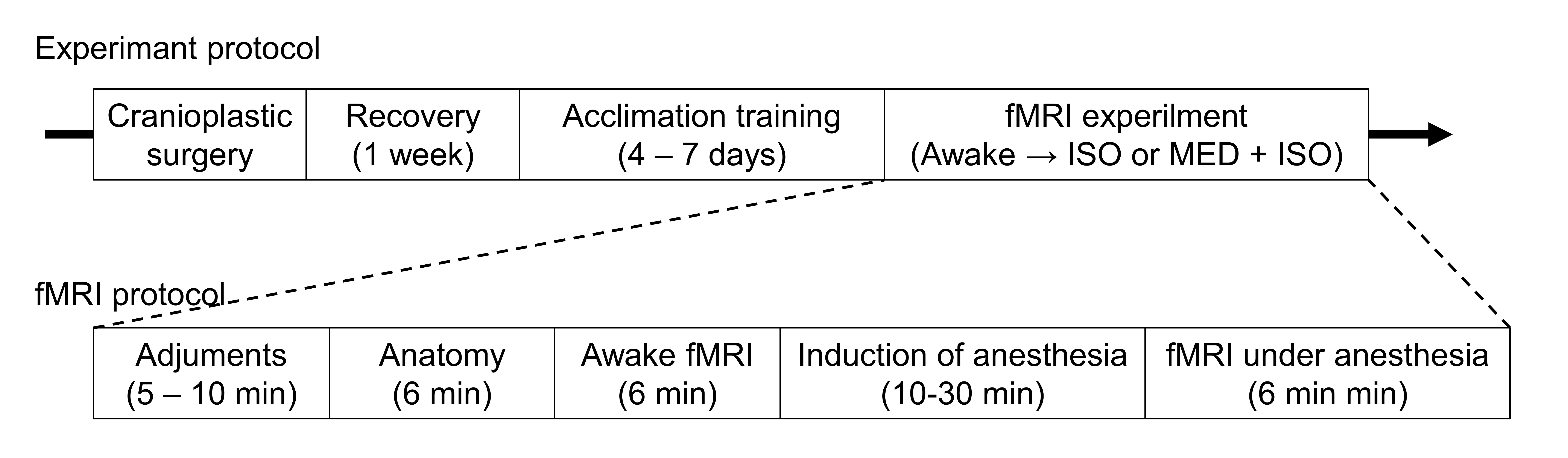
Figure 1 Schematic figure of the experimental protocol.
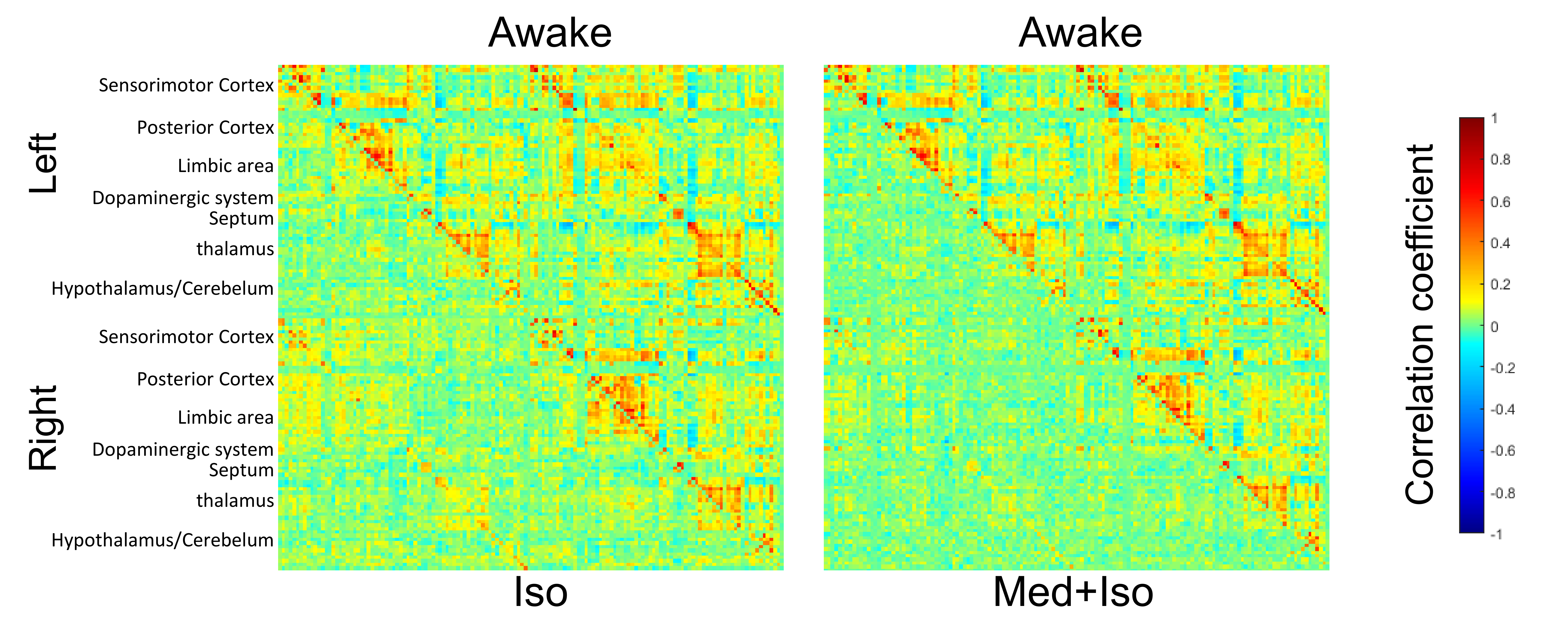
Figure 2 Averaged functional connectivity matrices under (left panel) ISO anesthesia (lower left) and awake state (upper right), and (right panel) MED + ISO anesthesia (lower left) and awake state (upper right). Color bar represents the Pearson correlation coefficients.
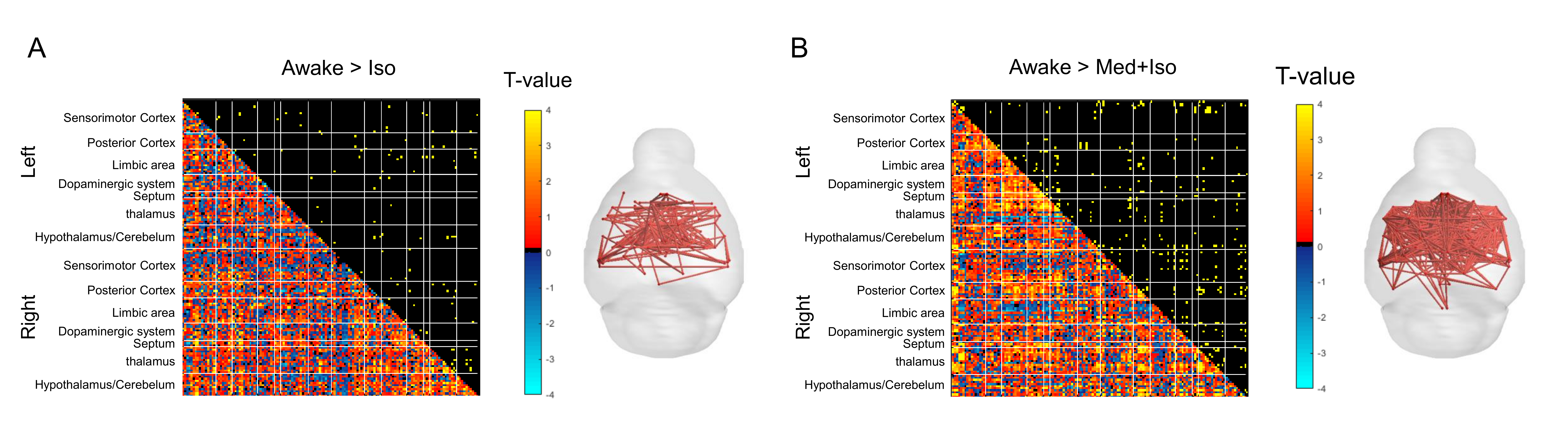
Figure 3 Significant changes of correlation coefficients (A) under ISO anesthesia and (B) under MED + ISO anesthesia compared with awake state. Right upper triangle panel shows the significant changes (p < 0.05, network-based statistic). Left lower panel shows the t-values. Figures are reconstructed significant changes in functional connectivity. Color bar shows t-values.
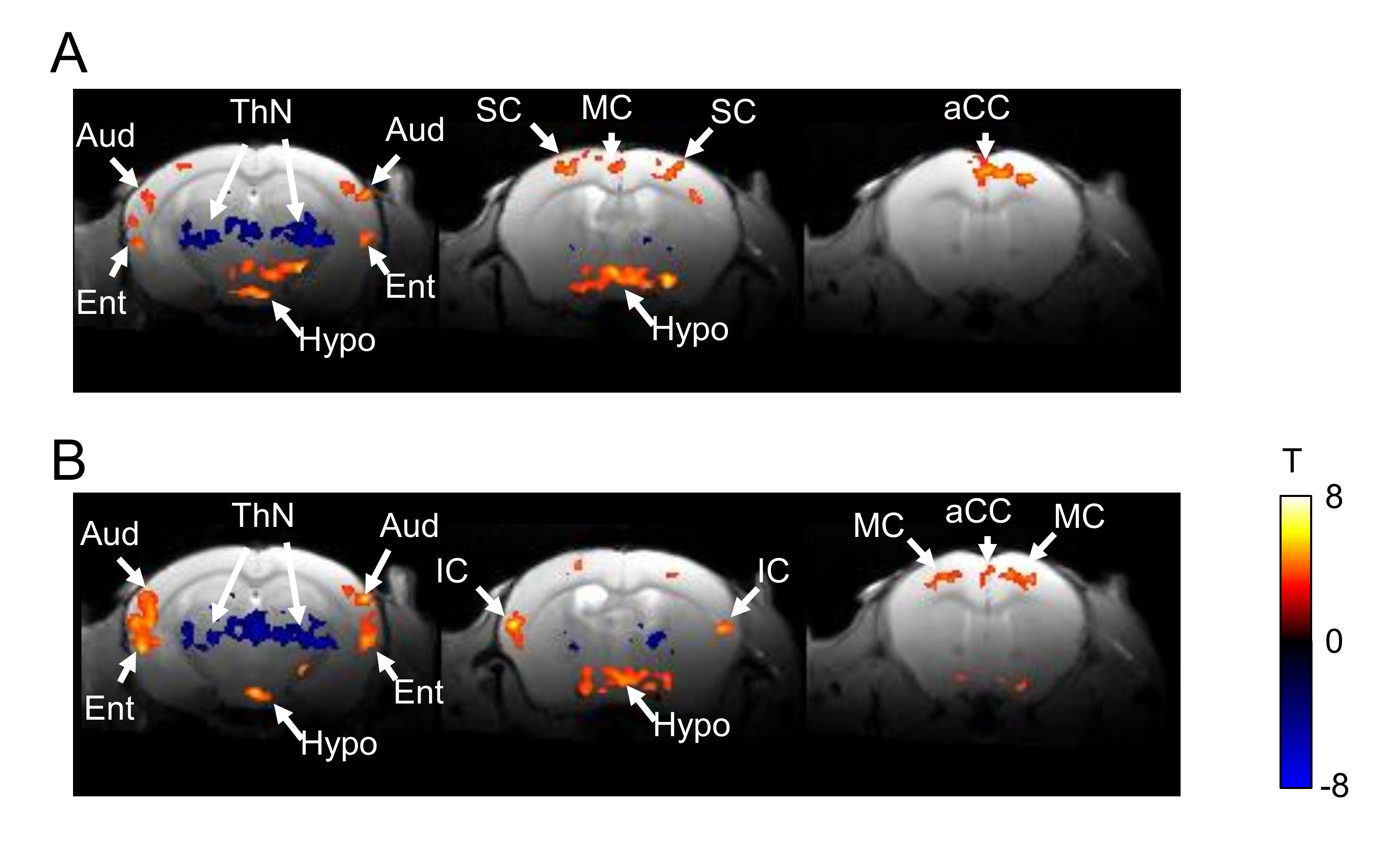
Figure 4 Significant increase or decrease of fALFF (A) under ISO anesthesia and (B) under MED + ISO anesthesia rather than awake state. Aud, auditory cortex; aCC, anterior cingulate cortex; Ent, entorhinal cortex; Hypo, hypothalamus; IC, insular cortex; MC, motor cortex; ThN, thalamic nuclei; SC, somatosensory cortex.