2921
Combined RS-fMRI and calcium recordings show stabile brain states in mice after switching anesthetic regimen1Department of Clinical Radiology, University Hospital Münster, Münster, Germany, 2Department of Anesthesiology, University Hospital Münster, Münster, Germany
Synopsis
Resting-state fMRI in mice became a popular method to investigate brain circuits. However, uncertainty resides over the dynamic effects of anesthetics on brain states and functional connectivity. Using a multimodal approach with optical calcium recordings and RS-fMRI, we assessed brain states and networks while switching anesthetic regimen from 1% isoflurane to a combined 0.2% isoflurane/medetomidine anesthesia. We find that brain states reach a steady state after a short transition time and detected that functional connectivity changes were strongest relative to the 1% isoflurane condition. We conclude that brain states and networks are stable from 30 minutes after switching anesthetic regimen.
INTRODUCTION
The popularity of resting-state (RS) functional (f)MRI is rising in preclinical research with still open questions on the effect of anesthetics on neuronal network dynamics. Recently, the combination of low-dose Isoflurane (ISO) and Medetomidin (MED) has become an advocated anesthetic regimen as it avoids respiratory depression and analgesic effects of high-dose ISO and MED, respectively1. Further, the opposing effects on vessel diameter are considered beneficial for fMRI studies. However, uncertainty resides over how fast relevant parameters adapt. Therefore, we investigated the brain state of mice during the transition from a single anesthetic (ISO) to a combined anesthetic regimen (ISO/MED) at different time points using a multimodal approach with RS-fMRI and calcium recordings.METHODS
Animals: 12 female C57/BL6J mice were used in this study to investigate brain states under different anesthetic conditions. MRI measurements were conducted as recently published2 on a 9.4 T Bruker Biospec 94/20 small animal scanner using a CryoProbe (Bruker Biospin). Functional MR acquisitions were recorded with a GE-EPI sequence (TR/TE:1000/18ms, 18 slices, 0.5mm slice thickness, FOV 28x26mm2, Matrix 80x80, 600 repetitions). RS-fMRI scans were acquired at different time points and under different anesthetic conditions. The first scan was performed under 1% ISO anesthesia. Subsequently, MED infusion (0.1mg/kg bolus, 0.2mg/kg*h continuous infusion) was started and ISO was slowly reduced to 0.2% within 30 minutes. RS-fMRI scans were further performed at the following timepoints: 25 min (with 0.6% ISO), 45 min (with 0.2% ISO), and 100 min (with 0.2% ISO) following the MED bolus injection. Data analysis: A digital mouse brain atlas consisting of 188 brain regions was registered to each EPI volume in its native space. Functional connectivity was determined using the multi-seed region approach3. For each animal, the average timecourses of a seed region of a ROI were correlated with the timecourse of every voxel in the brain, resulting in animal-specific asymmetric correlation matrices. For group comparison, these correlation matrices were averaged, and significant correlations were calculated using a t-test (p<0.05). The weight of each edge corresponded to its correlation coefficient r and indicated the strength of the functional connection. Typical characteristics of complex networks, including clustering coefficient, small-world index, averaged path-length, and connectivity strength, were calculated using MagnAn 2.5 (BioCom). For calcium recordings, animals were injected with Oregon Green™ Bapta-1 (OGB) on a stereotactic frame as previously published1. An optic fiber (diameter 200µm) was positioned in the injection site and connected to a blue laser (488 nm, Saphire, Coherent) to deliver excitation light. Upon binding of calcium ions to OGB, OGB emits green light (indicative of neuronal activity), which was detected using the same optic fiber. In a few experiments, RS-fMRI and calcium recordings were performed simultaneously.RESULTS
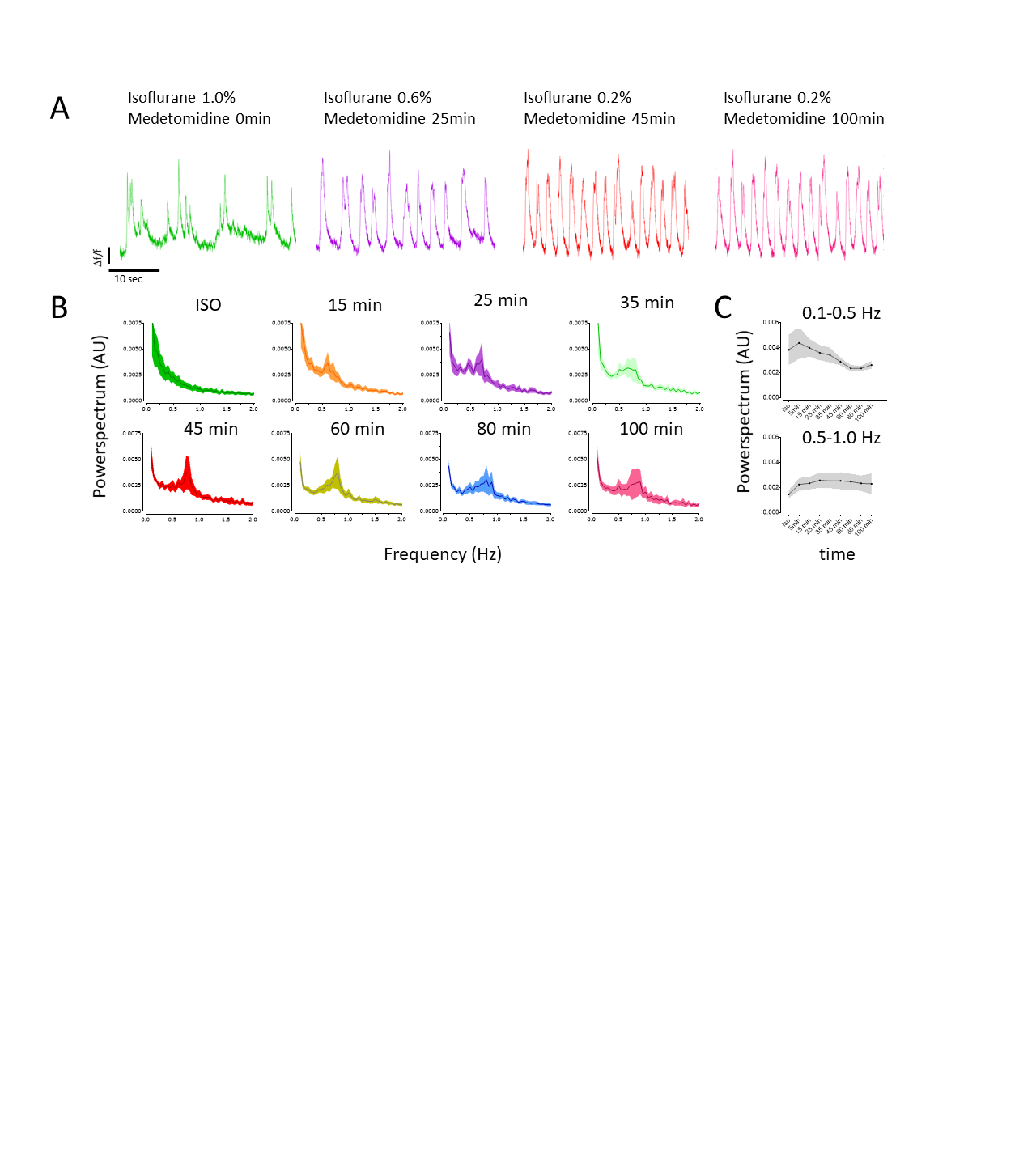
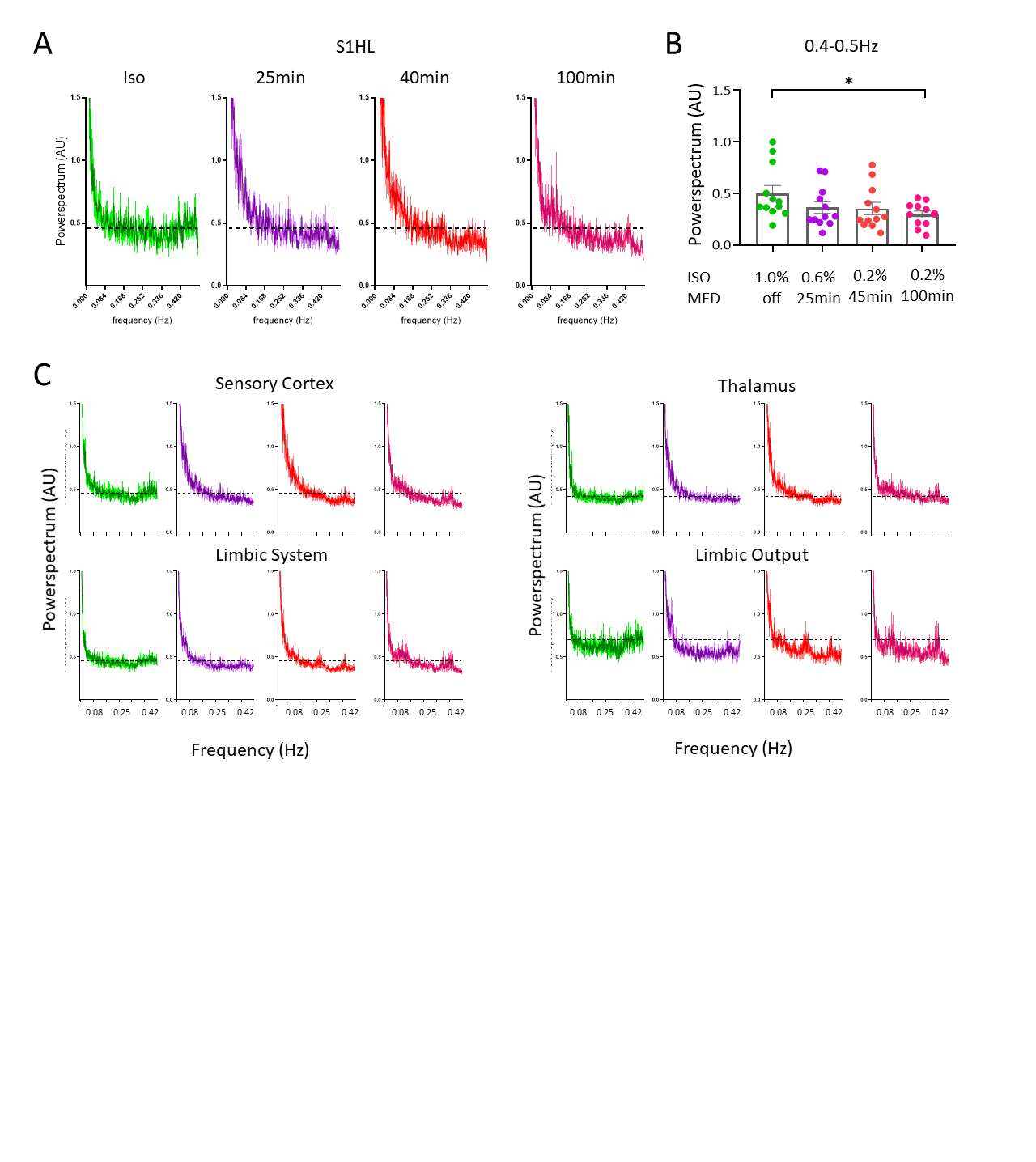
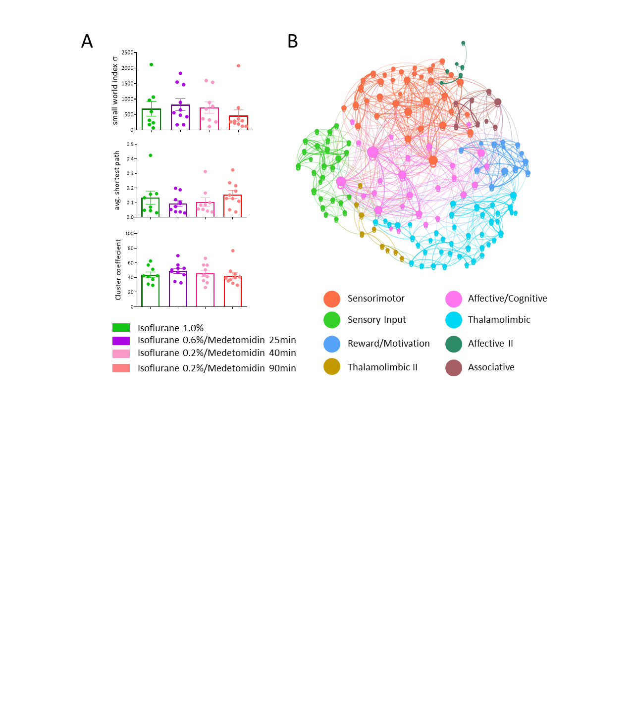
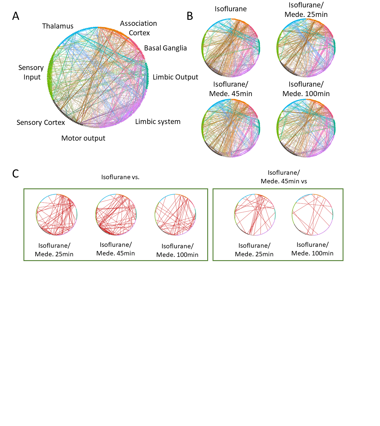
Calcium recordings and RS-fMRI consistently showed a change of brain state and network organization during the transition from ISO to ISO/MED sedation. Acute experiments with OGB show calcium transients at a frequency of <0.5Hz (1A, green) under ISO anesthesia. Within 5-25 minutes after the MED bolus injection, the frequency of calcium transients steadily increased to 0.5-1.0Hz (1A). From 35 min after MED bolus and with 0.2% ISO, the frequency of transients remained constant throughout the rest of the experiment (1A, 1C), which was accompanied by a decrease in low-frequency events (0.1-0.5Hz, 1C top). We then performed a Fourier transform of spontaneous BOLD activity in RS-fMRI data and a spectral analysis of different brain regions/functional anatomical groups (2A). Consistent with calcium recordings in S1HL we detected a decrease in low-frequency events throughout the change of anesthetic regimen that appeared to plateau within 25 to 45 minutes after MED bolus (2B). A similar effect was observed in different functional groups like the sensory cortex, limbic system (including hippocampus), limbic output regions (including hypothalamus), and thalamic regions (2C). We next calculated the global properties of RS-networks using graph theory. Measures for network segregation (clustering coefficient), normalized avg. path length and processing efficiency (small world index) did not show significant changes between different anesthetic conditions (3A). We detected eight communities with four major components that could be identified as sensorimotor, thalamolimbic, affective/cognitive, and sensory input (3B). Next, we investigated the effect of anesthesia on functional connectivity. The averaged functional correlations are displayed for each group (Fig.4B). Statistical analysis using a t-test (p<0.05) corrected for the same network density showed most significantly altered components between ISO and ISO/MED timepoints (ISO/MED25min: 39, ISO/MED 45min: 56, ISO/MED 100min: 28, Fig.4C, left panel). Network alterations between ISO/MED timepoints were less prominent and showed only 27 and 12 significantly altered components between ISO/MED 45min and ISO/MED 25min and ISO/MED 100 min, respectively.DISCUSSION
Calcium and RS-fMRI recordings clearly show brain states with slow-wave activity (UP-DOWN transitions) under ISO that rapidly changes to a persistent state upon switching the anesthetic regimen to ISO/MED in the primary sensory cortex. These changes were paralleled by alterations in the pattern of functional connectivity. Specifically, the coupling between the sensory cortex, association cortex, and the limbic system changed during the transition period.CONCLUSION
We recommend a 30-minute waiting period after switching from a single anesthetic regimen with ISO to combined ISO/MED anesthesia when studying the somatosensory cortex with RS-fMRI in mice.Acknowledgements
No acknowledgement found.References
1. van Alst, T. M. et al. Anesthesia differentially modulates neuronal and vascular contributions to the BOLD signal. Neuroimage 195, 89–103 (2019).
2. Albers, F., Wachsmuth, L., van Alst, T. M. & Faber, C. Multimodal Functional Neuroimaging by Simultaneous BOLD fMRI and Fiber-Optic Calcium Recordings and Optogenetic Control. Mol. Imaging Biol. 20, 171–182 (2018).
3. Kreitz, S., Alonso, B. de C., Uder, M. & Hess, A. A new analysis of resting state connectivity and graph theory reveals distinctive short-term modulations due to whisker stimulation in rats. Front. Neurosci. 12, 1–19 (2018).
Figures