2886
Feasibility of high resolution low-field MRI of balloon angioplasty in ex vivo porcine arteries with realistic blood flow
Eline Huizing1, Richte C.L. Schuurmann2, Frank F.J. Simonis3, Çağdaş Ünlü1, Henri G.D. Leuvenink4, and Jean-Paul P.M. de Vries2
1Department of Surgery, Northwest Clinics, Alkmaar, Netherlands, 2Department of Surgery, University Medical Center Groningen, Groningen, Netherlands, 3University of Twente, Enschede, Netherlands, 4Surgical Research Laboratory, University Medical Center Groningen, Groningen, Netherlands
1Department of Surgery, Northwest Clinics, Alkmaar, Netherlands, 2Department of Surgery, University Medical Center Groningen, Groningen, Netherlands, 3University of Twente, Enschede, Netherlands, 4Surgical Research Laboratory, University Medical Center Groningen, Groningen, Netherlands
Synopsis
Optimal treatment parameters of balloon angioplasty for revascularization such as inflation time and pressure are subject to debate. Limited data exists because accurate assessment of vascular function is challenging and therefore the influence procedure parameters have on arterial wall damage is largely unknown. This study shows that portable MRI at 0.125x0.125x2 mm³ resolution can be used for assessment of diameter and wall thickness during balloon angioplasty in ex vivo porcine arteries. Even after deflation and removal of the balloon the arterial diameter remained enlarged; this was coupled to a decrease in wall thickness.
Introduction
Revascularization remains the cornerstone for limb salvage in patients with severe lower limb ischemia1. In recent years, the endovascular approach has become the first choice for revascularization2. Traditionally, endovascular procedures were performed using a plain balloon, but many specific devices are being developed to facilitate the procedure and aim for better outcomes3. Also, optimal treatment options such as inflation time and inflation pressure are subject of debate. There is, however, limited data that identifies the optimal endovascular strategy and technique4. Further, the influence that different procedure parameters have on arterial wall damage is still unknown, and accurate assessment of vascular function is challenging. This study describes the feasibility of a portable low-field MRI scanner for accurate real-time assessment of artery function (diameter change) during balloon angioplasty in ex-vivo arteries with realistic blood flow.Method
A setup was built with realistic flow (70 ml/min) of porcine blood through a porcine cadaver artery flowing at physiological temperatures through ex vivo bench-top model. A 0.4T portable MRI scanner with 1 cm diameter bore (Pure Devices, Rimpar, Germany) was used for imaging. 2D MR scans were performed in transversal direction and longitudinal direction using a spoiled gradient echo sequence, with parameters as described in Table 1.Results
Scans were acquired before balloon insertion, after balloon inflation, after balloon deflation and finally after balloon removal; the resulting images are displayed in Figure 1. The acquired resolution of 0.125x0.125 mm² of the transversal scans is sufficient to accurately image the artery with an arterial wall thickness of 1.2 mm before inflation. The lumen diameter of the artery was 3.2 mm, which increased to 5.2 mm after balloon inflation. Because the balloon was filled with water, it can be clearly seen in the scans. After deflation of the balloon, the arterial lumen diameter remained increased compared to initial measurements (4.2 mm) and the wall thickness was decreased to 0.8 mm.Discussion
The resolution of the transversal and longitudinal low-field tabletop MRI images were sufficient to detect increase of the artery lumen diameter and decrease of the artery wall thickness. The boundaries between the balloon, lumen and vessel wall were clearly distinguishable when the vessel was placed inside the scanner. When the artery is surrounded by Dulbecco's Modified Eagle Medium (DMEM) instead of air, arterial function will be preserved. Subsequently, vessel injury can be assessed after balloon angioplasty using macroscopic and microscopic histopathology. However, since signal intensity of DMEM approaches that of the arterial wall, wall diameter assessment becomes more challenging. A solution would be to suppress the signal from DMEM by adding a high concentration of T2-shortening agent. The effect of these additives on the functioning of the artery has yet to be investigated. Currently, 2D analyses were performed with high spatial resolution and relatively low scan times, but 3D sequences are also optional, making isotropic resolution reachable. Future experiments will investigate the possibility of measuring luminal flow rate using the tabletop MRI.Conclusion
It is feasible to image the arterial wall of ex vivo porcine arteries using a portable low-field MRI scanner during several steps in a catheter inflation procedure. This enables measuring diameter and wall thickness change, together with vascular function this is essential for defining the optimal balloon angioplasty strategy.Acknowledgements
No acknowledgement found.References
1. Conte MS, Bradbury AW, Kolh P, et al. Global vascular guidelines on the management of chronic limb-threatening ischemia. J Vasc Surg. 2019;69(6S):3S-125S.e40.2. Aboyans V, Ricco J-B, Bartelink M-LEL, et al. Editor’s Choice - 2017 ESC Guidelines on the Diagnosis and Treatment of Peripheral Arterial Diseases, in collaboration with the European Society for Vascular Surgery (ESVS). Eur J Vasc Endovasc Surg Off J Eur Soc Vasc Surg. 2018;55(3):305-368.
3. Zeller T, Rastan A, Macharzina R, et al. Novel Approaches to the Management of Advanced Peripheral Artery Disease: Perspectives on Drug-Coated Balloons, Drug-Eluting Stents, and Bioresorbable Scaffolds. Curr Cardiol Rep. 2015;17(9):624.
4. Farber A, Eberhardt RT. The Current State of Critical Limb Ischemia: A Systematic Review. JAMA Surg. 2016;151(11):1070-1077.
Figures
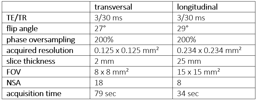
Table 1: MRI scan
parameters of the spoiled gradient echo sequence
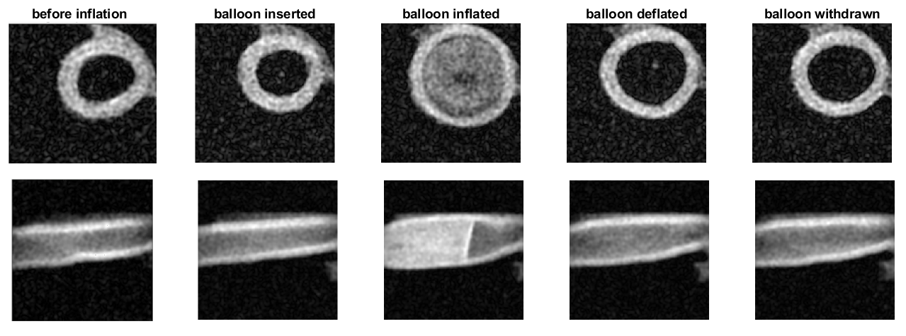
Figure 1: Transversal
(top row) and longitudinal (bottom row) slices through the artery showing the arterial wall and lumen
diameter in several steps during the procedure of balloon catheter inflation.