2879
Rapid Time-Resolved 4D MRA of the Brain Using Golden-Angle Radial Sparse Parallel (GRASP) MRI1Department of Radiology, Children's Healthcare of Atlanta, Atlanta, GA, United States, 2Department of Radiology, New York University Langone Medical Center, New York, NY, United States, 3Department of Neurology, New York University Langone Medical Center, New York, NY, United States
Synopsis
GRASP is a fast and flexible MRI technique combining compressed sensing, parallel imaging, and golden-angle radial sampling. GRASP MRI obtained as part of routine clinical care without specific angiographic parameters can be displayed as 4D MRA images with high spatial and temporal resolution. It exhibits excellent performance and high reader confidence in dynamic angiography, using a highly flexible plug-and-play reconstruction and visualization pipeline. Further exploration of diagnostic accuracy in disease-specific applications is warranted.
Introduction:
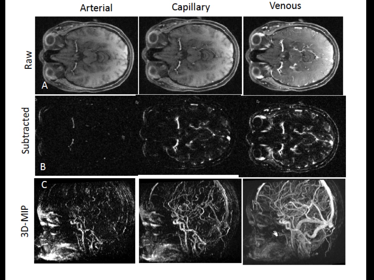
GRASP is a fast and flexible MRI technique combining compressed sensing, parallel imaging, and golden-angle radial sampling. It enables continuous data acquisition with retrospective user-defined reconstruction parameters optimized for application-specific use, e.g. monitoring of dynamic bolus passage with high temporal resolution.1 Herein, we explore the GRASP methodology using a custom reconstruction and visualization pipeline allowing multiplanar, subtracted, and time-resolved volumetric time series to isolate reconstructed brain MR angiography from anatomic brain imaging obtained in routine practice. The feasibility and performance of 4D GRASP MRA were examined in a three-rater study of angiographic quality and accuracy.Methods:
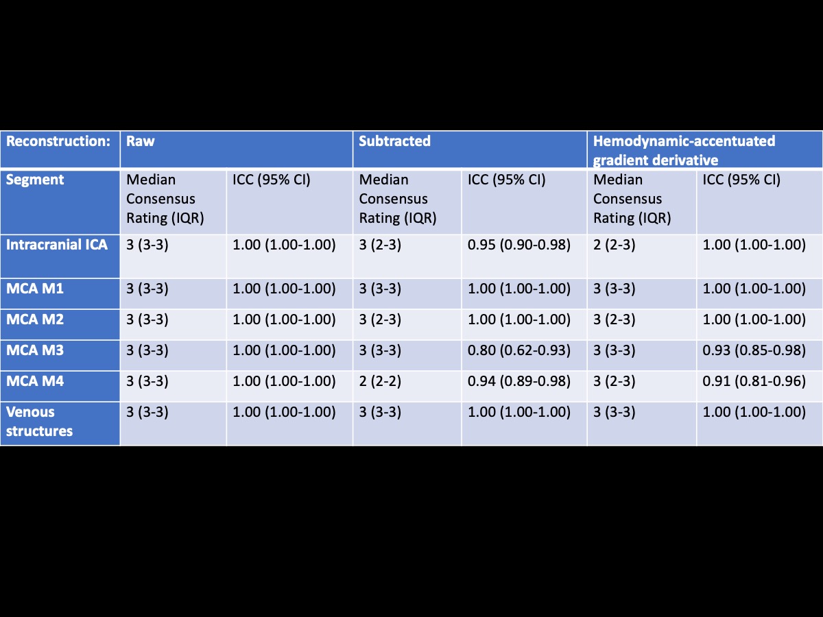
Axially-acquired GRASP scans obtained during the course of routine clinical care at 3T (MAGNETOM Skyra/Prisma, Siemens Healthcare, Germany) were reconstructed using a custom C++ implementation of the GRASP algorithm and post-processed with the MeVisLab software platform (MeVis Medical Solutions AG, Germany). A weight-based gadolinium dosing regime was employed in all subjects using a high-relaxivity agent (Gadavist) that was injected 20sec after the start of the GRASP acquisition. Images were reconstructed by combining 3 consecutive radial views into one frame, resulting in a time resolution of ~900ms/frame and spacial resolution of approximately 1.0mm3. Eleven examinations were scored by three neuroradiologists for image quality, dynamic imaging capabilities, and apparent imaging and angiographic reconstruction artifacts. Peak arterial and venous enhancement phases were identified and subsequently scored qualitatively using a 4-point Likert scale, with attention to 6 vascular segments: 1) intracranial ICA; 2) MCA M1; 3) MCA M2; 4) MCA M3; 4) MCA M4; 5) deep cerebral veins; and 6) dural venous sinuses. Raw images, subtracted angiography, and a hemodynamically accentuated sliding-frame, intensity-based gradient derivative dataset were made available to readers.Results:
Averaged across readers, a distinct arterial phase was identified in all 4D MRA reconstructions, with a median of 2 distinct frames (IQR1-3; Table 1). A capillary phase was identified in all subtractions and processed sequences, median of 2 distinct frames (IQR 2-3). Median Likert scale rating for vascular segments were 3 (excellent, diagnostic quality) for all vascular segments between the three readers for all reconstructions, with excellent interclass correlation (1.00-0.93). Overall, there were few artifacts identified.Discussion:
4D MRA using GRASP exhibits excellent performance and high reader confidence in dynamic angiography, using a highly flexible plug-and-play reconstruction and visualization pipeline. While existing 4D dynamic MRA techniques have been previously reported, several attributes condition GRASP uniquely for optimized and flexible performance by comparison with past approaches. Because GRASP uses a continuous radial stack-of-stars acquisition of k-space, scans do not depend on exact timing of the data acquisition, which significantly simplifies the examination in routine clinical practice. Moreover, an arbitrary number of consecutive radial views can be combined into a single image frame to create angiographic imaging of any desired temporal resolution. An additional advantage to radial k-space sampling is its reduced sensitivity to motion artifacts. Importantly, GRASP simultaneously provides anatomical T1-weighted images with high spatial resolution as well as dynamic angiographic images of the entire imaging volume, allowing for the assessment of tissue arteriovenous shunting by identifying differences in parenchymal capillary phases. This altogether makes GRASP an intriguing and attractive approach for dynamic MRA.Conclusion:
GRASP MRI obtained as part of routine clinical care without specific angiographic parameters can be displayed as 4D MRA images with high spatial and temporal resolution. Further exploration of diagnostic accuracy in disease-specific applications is warranted.Acknowledgements
No acknowledgement found.References
1. Feng L, Grimm R, Block KT, et al. Golden-angle radial sparse parallel MRI: combination of compressed sensing, parallel imaging, and golden-angle radial sampling for fast and flexible dynamic volumetric MRI. Magn Reson Med 2014;72:707-717
2. Chandarana H, Feng L, Block TK, et al. Free-breathing contrast-enhanced multiphase MRI of the liver using a combination of compressed sensing, parallel imaging, and golden-angle radial sampling. Invest Radiol 2013;48:10-16
Figures