2875
Deep Learning Algorithms May Aid In The Evaluation Of Cine Images In Patients With Atrial Arrhythmia – A Case Series1Cardiovascular imaging, RI-McGill University Health Center, Montreal, QC, Canada, 2Faculty of Medicine, McGill University Health Center, Montreal, QC, Canada, 3RI-McGill University Health Center, Montreal, QC, Canada, 4Faculty of Medicine, University of Alberta, Edmonton, Alberta, AB, Canada, 5MR applications and workflow, GE Healthcare, Montreal, QC, Canada
Synopsis
Cardiovascular magnetic resonance cine imaging is utilised to give comprehensive information on ventricular function. Arrhythmia obstructs acquisition of these images, where the use of faster acquisition protocols with deep learning reconstruction methods may aid in solving the problem. We evaluated cine images of three patients in atrial arrhythmia, acquired using standard method; fast, variable density spatiotemporal sampling acquisition (VD kt) of one(1rr) or three(3rr) heart beats; and deep learning reconstruction of the same (DL-1rr,DL-3rr). Our results showed that undersampled techniques combined with deep-learning algorithms result in image quality improvements with no significant difference in quantitative values between all acquisition techniques.
Introduction:
Cine images in cardiovascular magnetic resonance imaging are the gold-standard to quantify and give comprehensive information of the ventricular function. ECG gating is usually employed to acquire cine images; however, arrhythmia obstructs ECG gating and results in poor image quality. To overcome these issues, faster acquisition protocols with DL reconstruction methods are being used.Methods:
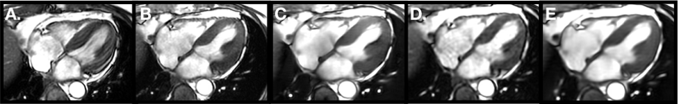
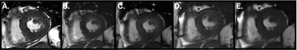
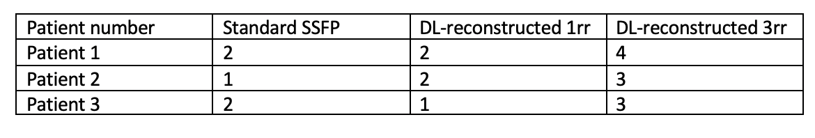
Three patients with a history of atrial fibrillation/flutter, awaiting ablation for the arrhythmia, were referred for a Cardiac MRI (CMR) at different times, for evaluation of left and right ventricle (LV, RV) function and pulmonary vein anatomy. Each of them had atrial fibrillation during the CMR scan. Cine images were acquired using standard ASSET-accelerated SSFP (FIESTA) cine sequence (R=2); a fast, variable density spatiotemporal sampling acquisition (VD kt) of one (1rr, R=12) or three (3rr, R=4) heart beats, respectively. The VD kt images were reconstructed using kt-ARC or using a Deep Learning (DL) ESPIRiT algorithm(1). The images were analyzed by a blinded, experienced reader, who scored images from 0-4 (see Figure 3).Results:
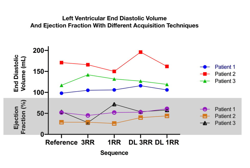
Images acquired using standard cine had the worst image quality (mean 1.67), and the visual analysis and volumetric quantification of LV and RV functional parameters was considered unreliable. Conversely, the DL-reconstructed images acquired in 3rr had a significantly better image quality (mean 3.33) (Figure 3). The quantitative evaluation using standard SSFP, kt-ARC 1rr, kt-ARC 3rr, DL reconstructed 1rr and 3rr resulted in values which showed no significant difference (Figure 4).Discussion:
In patients with atrial fibrillation, deep-learning based image quality improvement algorithms can improve the subjective image quality even when applied to undersampled images. While these observations indicate a significantly improved image quality for diagnosis in these patients, these results however do not prove that a subjectively better image quality also improves the accuracy of quantitative results.Conclusion:
Undersampled techniques combined with deep-learning informed algorithms results in image quality improvements which may be useful in patients with arrhythmia, who otherwise may require longer scan times and unreliable diagnostic image quality.Acknowledgements
No acknowledgement found.References
1. Sandino CM, Lai P, Vasanawala SS, Cheng JY. Accelerating cardiac cine MRI using a deep learning‐based ESPIRiT reconstruction. Magn Reson Med [Internet]. 2021 Jan 22 [cited 2020 Dec 13];85(1):152–67. Available from: https://onlinelibrary.wiley.com/doi/10.1002/mrm.28420Figures