2861
UNIform COmbined RecoNstruction (UNICORN) for 7T Clinical Fat-Suppressed TSE Imaging of the Human Knee1Siemens Medical Solutions USA Inc., Houston, TX, United States, 2Siemens Medical Solutions USA Inc., Rochester, MN, United States, 3Department of Radiology, Houston Methodist Research Institute, Houston, TX, United States
Synopsis
Fat-suppressed turbo-spin-echo (TSE) imaging is very important to visualize knee pathology such as cartilage defects in clinical routine. Ultra-high-field 7T magnetic resonance imaging (MRI) provides higher signal-to-noise-ratio (SNR) and contrast-to-noise-ratio (CNR) than 3T and 1.5T MRI, enabling better visualization of fine anatomical structures and physiological effects. However, imaging at 7T has higher receive and transmit non-uniformity that may degrade its clinical value. Recently, a UNIform COmbined RecoNstruction (UNICORN) algorithm was proposed for reducing the receive non-uniformity. The purpose of this preliminary work is to quantitatively and qualitatively evaluate and optimize UNICORN performance for 7T clinical TSE with fat suppression.
INTRODUCTION:
Ultra-high-field 7T magnetic resonance imaging (MRI) provides higher signal-to-noise-ratio (SNR) and contrast-to-noise-ratio (CNR) than 3T and 1.5T MRI, enabling better visualization of fine anatomical structures and physiological effects. Imaging at 7T, however, has higher receive and transmit non-uniformity that may degrade its clinical value. Recently, a UNIform COmbined RecoNstruction (UNICORN) algorithm was proposed for reducing the receive non-uniformity1. In that study, three radiologists qualitatively evaluated UNICORN performance on non-fat suppressed 7T turbo-spin-echo (TSE) knee MR images for SNR, uniformity, contrast, and overall image quality. The three radiologists preferred UNICORN with high inter-rater-agreement for uniformity, contrast, and overall image quality. The inter-rater agreement regarding SNR was moderate, possibly because the non-fat suppressed 7T TSE images had high enough SNR. Because fat suppression is very important in clinical knee imaging to enhance the visibility of pathology such as cartilage defects, the purpose of this preliminary work is to quantitatively and qualitatively evaluate and optimize UNICORN performance for 7T clinical TSE with fat suppression.METHODS:
Data Acquisition:This work was conducted as part of an institutional review board approved clinical trial performed on a MAGNETOM Terra scanner (Siemens Healthcare, Erlangen, Germany) using a single-channel transmit, 28-channel phased-array receive knee coil (QED, Quality Electrodynamics, Mayfield Village, OH). One subject’s knee was imaged in a sagittal orientation using a two-dimensional TSE sequence with a refocusing flip angle (FA) of 126o. The scan was repeated with a refocusing FA of 163o, keeping all other scan parameters identical. The acquisition parameters included: field-of-view (FOV) = 160 x 144 mm2, in-plane resolution = 0.4 x 0.4 mm2, slice thickness = 2 mm, number of slices = 47, echo time (TE) = 31 ms, repetition time (TR) = 6000 ms, spectral fat suppression ON, generalized auto-calibrating partially parallel acquisitions (GRAPPA) acceleration factor = 2. In addition, the transmit-field (B1) filter was turned ON to reduce the B1-induced non-uniformity2. Scan time for each acquisition was 4:50 min.
Image Reconstruction:
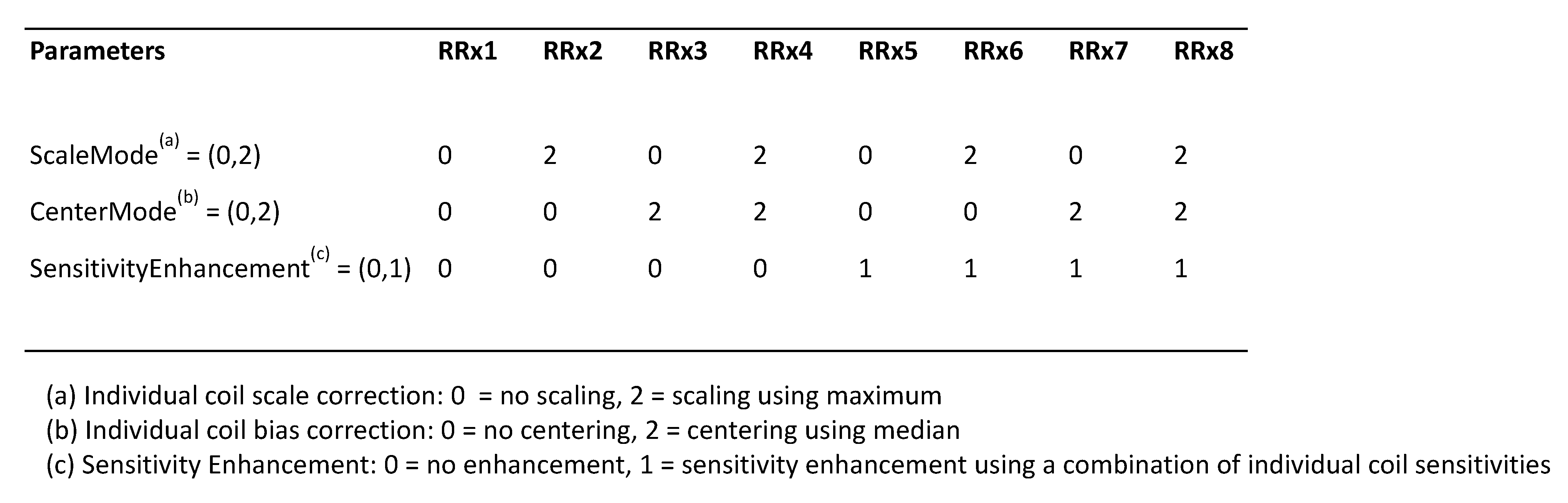
The raw measurement data was retrospectively reconstructed using a prototype implementation of the UNICORN algorithm with eight different parameter settings denoted as RRx1 to RRx8 (Table 1). Each setting reconstructed three image series per acquisition: no normalization (original), with UNICORN normalization (UNICORN), and with both UNICORN and B1 normalization (UNICORN+B1). There were 48 total reconstructed image series.
Quantitative Image Analysis:
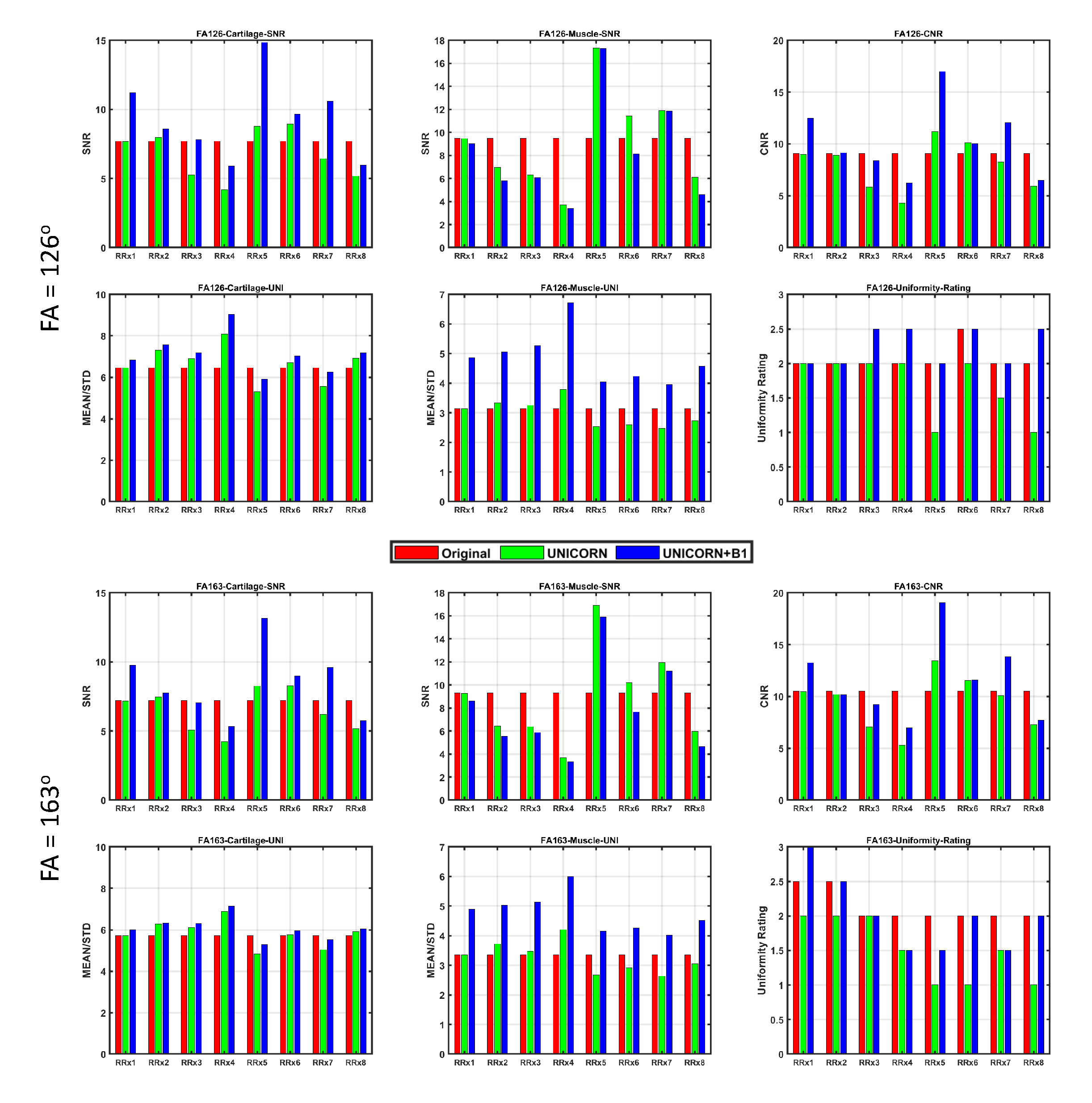
Quantitative image analysis was performed in MATLAB (MathWorks, Natick, MA). A board-certified body radiologist manually drew five regions of interest (ROIs) on the original images: one large region of cartilage (number of voxels = 478), one large region of skeletal muscle (number of voxels = 9031), two adjacent small regions of cartilage (number of voxels = 207) and synovial fluid (number of voxels = 121), and one large region of background for noise estimation (number of voxels = 814). The SNR of the two large muscle and cartilage ROIs was defined as the mean voxel intensity of each ROI divided by the mean voxel intensity of the noise ROI. A metric of uniformity, UNI, was defined as the mean voxel intensity of the two large ROIs divided by the standard deviation of the voxel intensities within the same ROI. CNR was defined as the difference between the mean voxel intensities of the small cartilage and fluid ROIs divided by the mean voxel intensity of the noise ROI.
Qualitative Clinical Evaluation:
The radiologist, who was blinded to the quantitative ROI analysis, also rated each image series on a 0-3 scale (0 = worst, 3 = best) in terms of SNR, uniformity, contrast.
RESULTS:
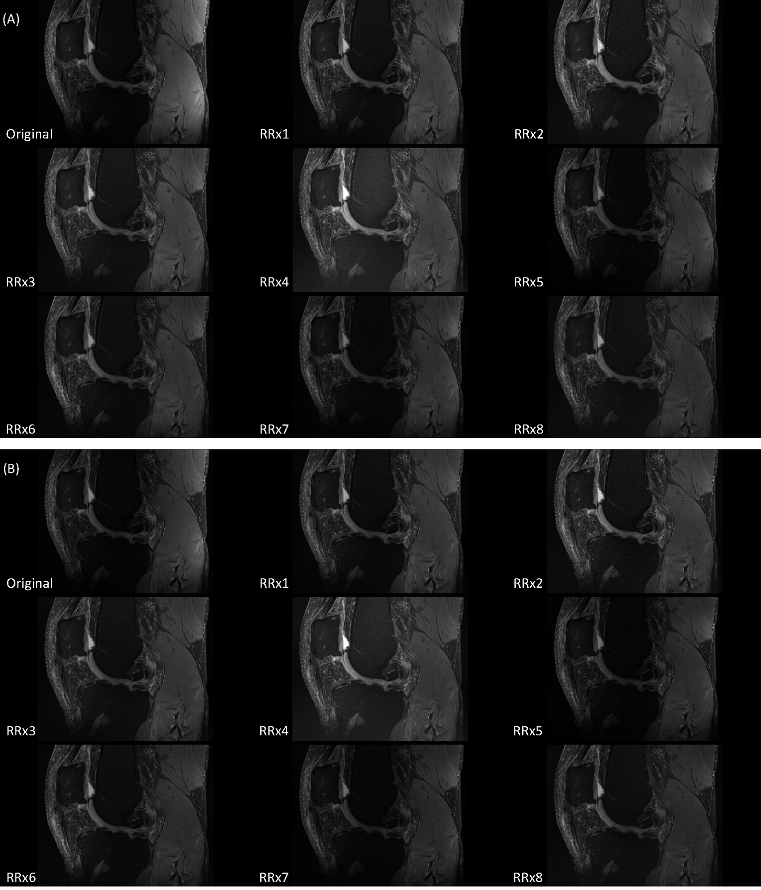
As shown in Figure 1, the UNICORN+B1 images of RRx4 (ScaleMode = 2, CenterMode = 2, and SensitivityEnhancement = 0) had the highest UNI of the large cartilage and muscle ROIs, while the UNICORN+B1 images of RRx5 (ScaleMode = 0, CenterMode = 0, and SensitivityEnhancement = 1) had the highest SNR of these two ROIs as well as the highest CNR. Exemplary images of the original and UNICORN+B1 images of RRx1 to RRx8 are displayed in Figure 2. Clinical evaluation showed that all the settings had very good SNR (all ratings = 3) and contrast (all ratings = 3). Compared with the original images, the UNICORN+B1 images of all the settings had the same or better uniformity (Fig. 1), consistent with the findings from the quantitative analysis.DISCUSSION:
This preliminary work demonstrates that UNICORN plus B1 filtering is a promising reconstruction tool to improve image quality of clinical fat-suppressed TSE of the knee at 7T. The limitations of this work are small sample size and lack of inter-rater validation. Future work will include more subjects and more ratersCONCLUSION:
UNICORN is a promising method to improve uniformity and contrast of 7T clinical fat-suppressed TSE images of the human knee.Acknowledgements
This work is supported by Methodist - Siemens Collaborative Research Funding.References
1. Chebrolu VV, Kollasch PD, Deshpande V, Grinstead J, Howe BM, Frick MA, Fagan AJ, Benner T, Heidemann RM, Felmlee JP, Amrami KK. Uniform combined reconstruction of multichannel 7T knee MRI receive coil data without the use of a reference scan. J Magn Reson Imaging. 2019 Nov;50(5):1534-1544. doi: 10.1002/jmri.26691. Epub 2019 Feb 19. PMID: 30779475.
2. Jellus, V., and B. Kiefer. "Optimization of the homomorphic filter for bias field correction." In Proc Intl Soc Magn Reson Med., 2005, Vol. 13, p.2247.
Figures