2792
Greater increase in magnetic susceptibility following acute MS lesion formation is associated with reduced myelin repair1Department of Neurology, Weill Cornell Medicine, New York, NY, United States, 2Department of Radiology, Weill Cornell Medicine, New York, NY, United States, 3Department of Population Health Sciences, Weill Cornell Medicine, New York, NY, United States
Synopsis
Quantitative susceptibility mapping (QSM) provides an effective means to directly map the distribution of magnetic susceptibility sources, such as brain iron. In this study, we examined the relationship between initial QSM change and short-term myelin recovery, measured by myelin water fraction (MWF). The results showed that change in lesion susceptibility at three months was significantly and negatively associated with change in MWF at one year. This significant association provides evidence of the damaging effect of brain iron during early stages of lesion development.
Introduction
Gradient echo (GRE) sequences are sensitive to iron1 and have been used to explore iron dynamics in multiple sclerosis (MS)2. Quantitative susceptibility mapping (QSM) provides an effective means to directly map the distribution of magnetic susceptibility sources including brain iron by solving the field-to-source dipole inversion problem3. Previous studies have showed that lesion susceptibility, as measured by QSM, increases rapidly following the formation of an acute Gadolinium-enhancing lesion and can be related to both myelin breakdown and iron-mediated inflammation4,5. We postulate that early iron accrual contributes to the toxic milieu of the lesion, which promotes tissue destruction and impedes myelin repair. In this longitudinal study, we examined the relationship between early QSM susceptibility change (initial three months) and short-term lesion myelin recovery, as measured by myelin water fraction (MWF), one year after lesion enhancement.Methods
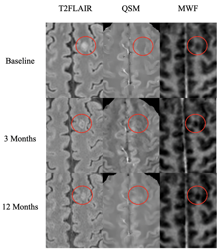
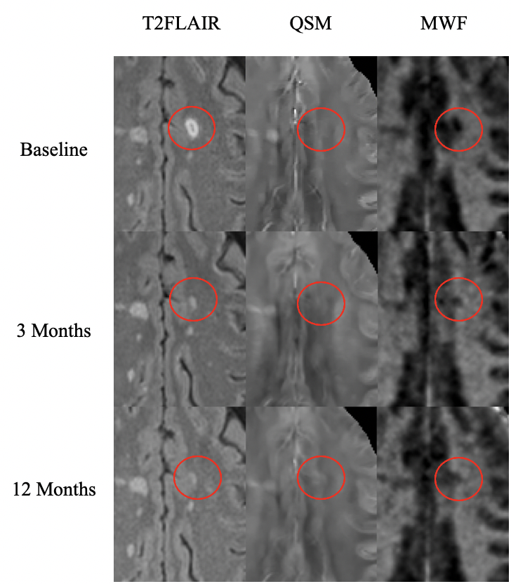
Thirteen MS patients had longitudinal QSM and MWF imaging in addition to conventional anatomical sequences at 3T (Siemens Skyra scanner). QSM (4.5 min) was reconstructed from complex GRE images using a fully automated Morphology Enabled Dipole Inversion (MEDI+0) method zero-referenced to the ventricular cerebrospinal fluid6. MWF (5 min) was acquired using Fast Acquisition with Spiral Trajectory and T2prep (FAST-T2) sequence and computer using three-pool non-linear least squares data fitting7. Each patient’s baseline scan was defined as the appearance of a new Gadolinium-enhancing lesion and longitudinal scans were performed at 3 months (± 30 days) and 12 months (+ 90 days). All images were co-registered to baseline QSM image space using FSL neuroimaging software. Lesion masks were traced on the co-registered T2FLAIR image and then overlaid on QSM, MWF, and contrast-enhanced T1 images. The susceptibility values were measured in reference to the adjacent normal appearing white matter (NAWM) to account for local fiber tract orientation. The relationship between the susceptibility change (baseline to three months) and myelin recovery (MWF change from baseline to one year) was modeled in a linear mixed effects model. Change in MWF was the model outcome and both change in susceptibility and lesion volume at baseline were considered as fixed effects. A random effect for subject was included to account for correlation in the data.Results
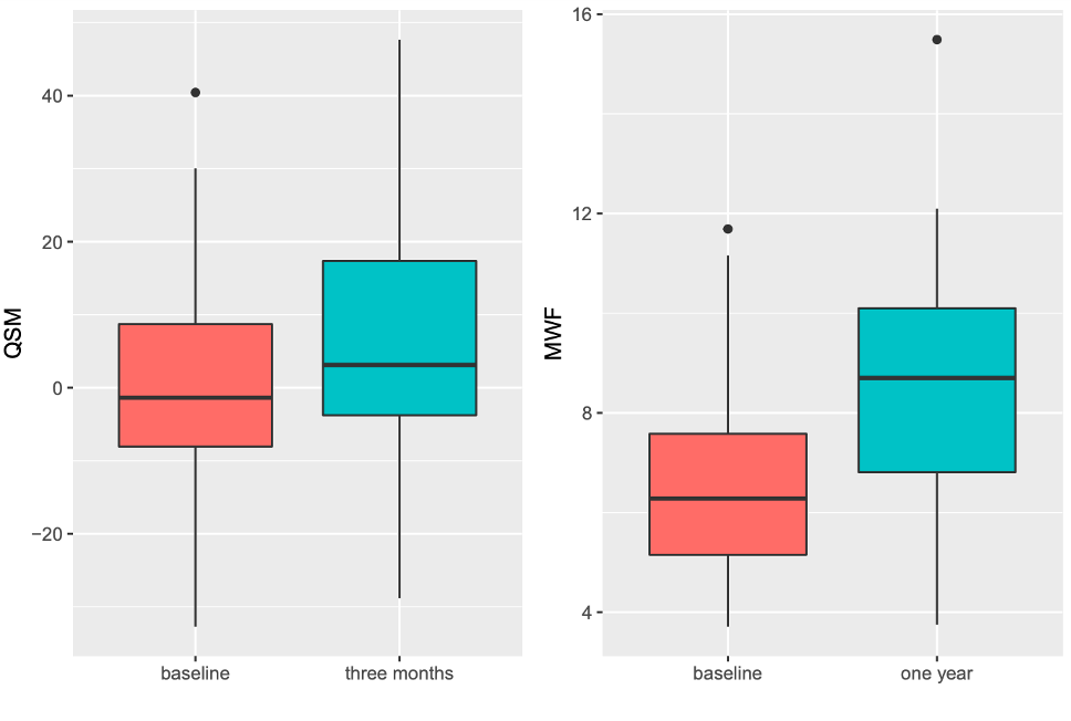
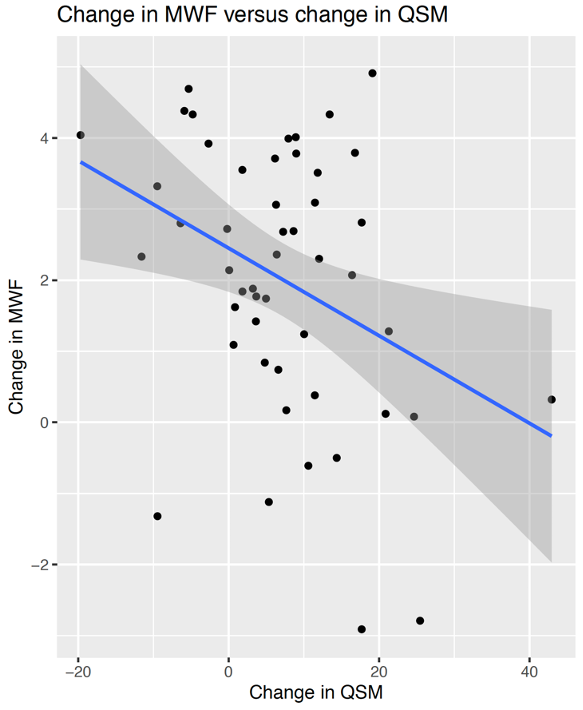
Forty-nine new Gadolinium-enhancing lesions were included in the analysis. At baseline, the average lesion volume was 461.23 mm3 ± 538.44, QSM susceptibility = 1.35 ppb ± 13.99, and MWF = 6.48% ±1.89. Lesion susceptibility increased significantly at three months to 7.11 ppb ± 10.9, p <0.001 (Fig. 3). Overall, there was an increase in MWF at 12 months, with a mean improvement of 2.01% ± 1.89, p <0.001 (Fig. 3). In the linear mixed effects model, after controlling for lesion volume (p=0.04), the change in susceptibility at three months was found to be negatively and significantly associated with MWF at one year (p = 0.039). This association is visually represented in Figure 1 and 2. The coefficient for the fixed effect of change in QSM is -0.04, which indicates that for a one-unit increase in change in QSM, on average, the MWF change decreases by 0.04 units (Fig. 4).Conclusion
In this longitudinal study of enhancing MS lesions, we found that a higher increase in susceptibility, occurring early after lesion development, is associated with less myelin recovery at one year. Given that lesion iron contributes to the rise in susceptibility, these results provide evidence of the damaging effect of iron release during early stages of lesion development and promote the use of QSM as a biomarker to further understand factors related to myelin recovery in acute MS lesions.Acknowledgements
No acknowledgement found.References
1. Langkammer C, Krebs N, Goessler W, Scheurer E, Ebner F, Yen K, et al. Quantitative MR imaging of brain iron: a postmortem validation study. Radiology. 2010;257(2):455-62.
2. Liu C, Li W, Tong KA, Yeom KW, Kuzminski S. Susceptibility-weighted imaging and quantitative susceptibility mapping in the brain. J Magn Reson Imaging. 2015;42(1):23-41.
3. Wang Y, Liu T. Quantitative susceptibility mapping (QSM): Decoding MRI data for a tissue magnetic biomarker. Magn Reson Med. 2015;73(1):82-101.
4. Zhang Y, Gauthier SA, Gupta A, Comunale J, Chia-Yi Chiang G, Zhou D, et al. Longitudinal change in magnetic susceptibility of new enhanced multiple sclerosis (MS) lesions measured on serial quantitative susceptibility mapping (QSM). J Magn Reson Imaging. 2016.
5. Deh K, Ponath GD, Molvi Z, Parel GT, Gillen KM, Zhang S, et al. Magnetic susceptibility increases as diamagnetic molecules breakdown: Myelin digestion during multiple sclerosis lesion formation contributes to increase on QSM. J Magn Reson Imaging. 2018;48(5):1281-7.
6. Liu Z, Spincemaille P, Yao Y, et al. MEDI+0: Morphology enabled dipole inversion with automatic uniform cerebrospinal fluid zero reference for quantitative susceptibility mapping. Magn Reson Med 2018;79:2795-2803
7. Nguyen TD, Deh K, Monohan E, Pandya S, Spincemaille P, Raj A, et al. Feasibility and reproducibility of whole brain myelin water mapping in 4 minutes using Fast Acquisition with Spiral Trajectory and adiabatic T2prep (FAST-T2) at 3 Tesla. Magn Reson Med. 2016;76(2):456-65.
Figures