2788
Computer-aided diagnosis of human breast lesion in T1 perfusion MRI using curvelet based features.1University of Pennsylvania, Philadelphia, PA, United States, 2, Indian Institute of Technology Delhi, New Delhi, India, Delhi, India
Synopsis
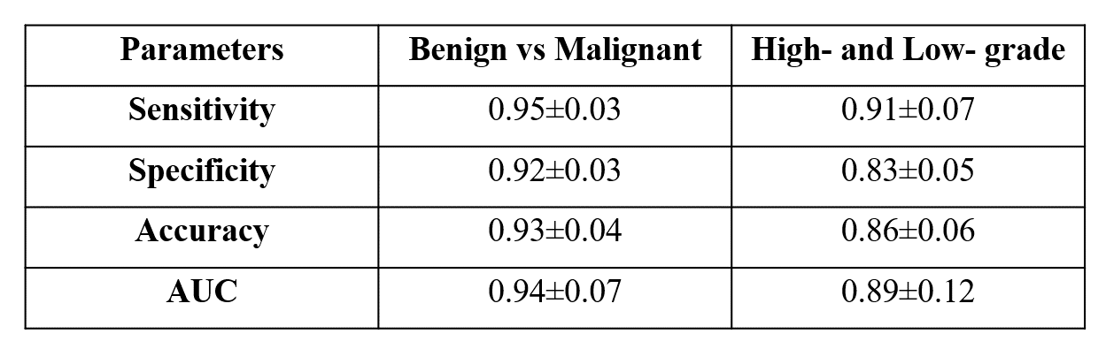
Curvelet transform is used as a multi-scale level decomposition to represent images. It was hypothesized that curvelet based texture features extraction can improve accuracy of tumor classification. The objective of this study was to differentiate the breast tumor using curvelet based features extraction followed by principal component analysis(PCA) for feature reduction and support vector machine(SVM) classifier. The study included T1 perfusion MRI data of 40 patients with breast cancer. The curvelet based texture feature using PCA with SVM classifier provided high average accuracy(0.93±0.04) in classification of malignant vs. benign and average accuracy (0.86±0.06) in characterization of high- vs. low-grade.
Introduction:
MRI is one of the widely used techniques in diagnosis and monitoring of treatment responses1. The histological grading provides important prognostic information in breast cancer2. Computer-assisted-diagnosis (CAD) systems of breast tumor in imaging has potential role towards its early diagnosis, which may significantly reduce the mortality rate and inter-observer variations in interpretations3. The characterization of breast lesions in T1 perfusion MRI provided varying sensitivity and specificity using different features such as morphology, texture, tracer kinetic, hemodynamic features, and etc. as reported previously4,5,6,7. Most of the reported studies are on differentiation between malignant vs. benign and quite less studies are on grading of tumors. Previously, curvelet transform8 has the ability to estimate the images in different decomposition levels and used for various image processing and computer vision applications such as image de-noising and reconstruction, and etc.9,10. It was hypothesized that texture features for each wedge in curvelet transform can improve accuracy of lesion classification in the current study. The purpose of this study was to develop a computer aided diagnosis for characterizing of breast lesion (benign vs. malignant and high-grade vs low-grade) using curvelet based features extraction and SVM classifier with 5-fold-cross validation.Methods:
All the MRI experiments were performed at 3T whole body Ingenia MRI system (Philips Healthcare, The Netherlands) using a 7 channel biopsy compatible breast coil. Forty female subjects (15 benign, and 25 with breast cancer) were scanned for MRI data.MRI Data acquisition: Fat saturation was based upon DIXON method. Dynamic 4D images with fat saturation were acquired using turbo spin echo pulse sequence. Multiple slices, covering entire breast tissue with slice thickness of 3 mm were acquired. FOV = 338 *338 mm2 and matrix size = 512 * 512 were used. T1 perfusion MRI was performed using a 3-dimensional fast field echo (3D-FFE) sequence (TR/TE = 3.0/1.5 ms, flip angle = 12 degree). Gd-BOPTA (Multihance, Bracco, Italy) in a dose of 0.1 mmol/kg body weights was administered intravenously with the help of a power injector at a rate of 3.0 mL/sec, followed by a bolus injection of a 30-mL saline flush. Forty time points were acquired with a temporal resolution approximately of 5.4 seconds for each time point.
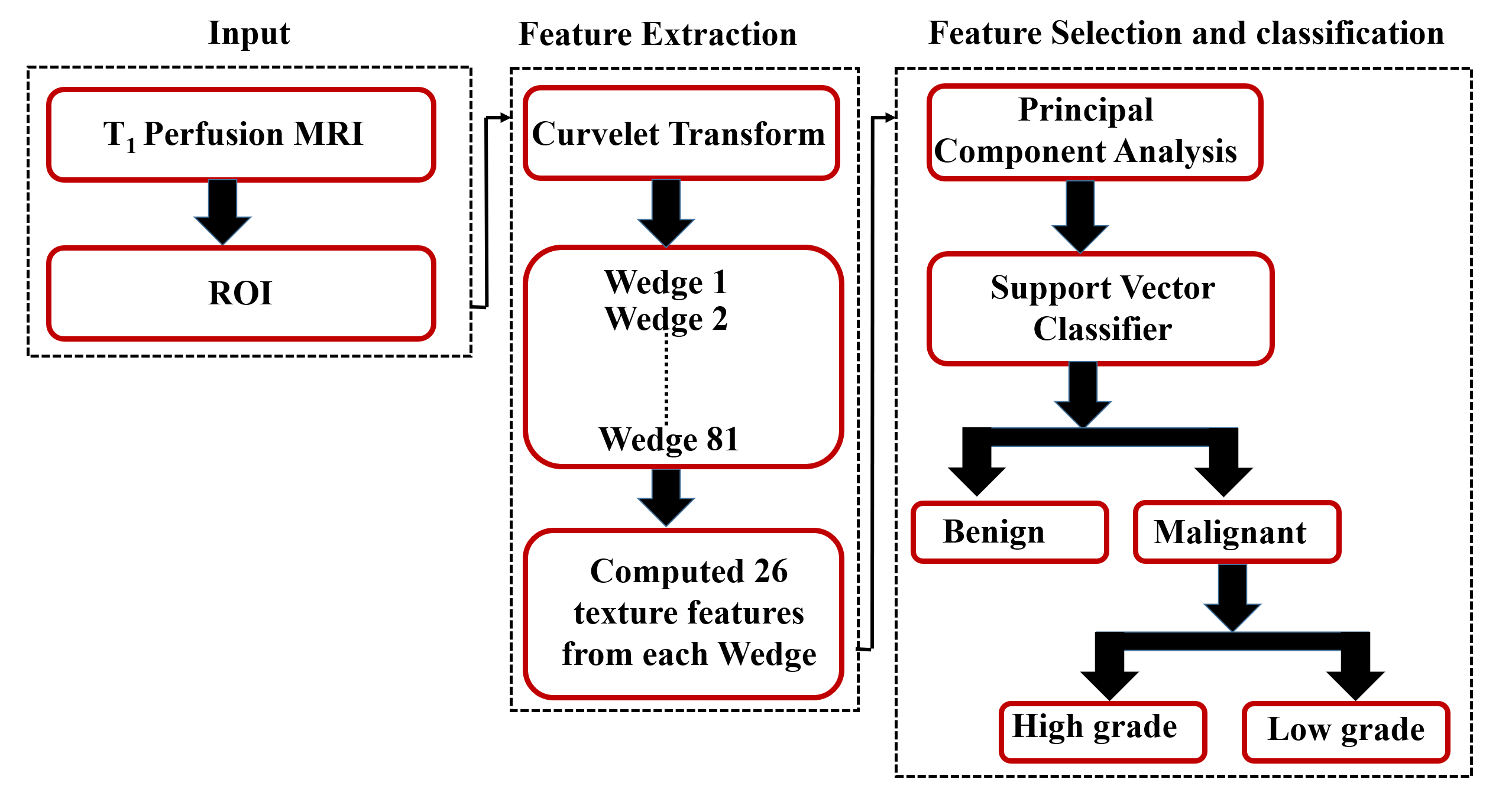
Data Processing: A systematic approach comprising of curvelet based feature extraction and classification was carried out as shown in Figure 1. The curvelet based texture features11 of regions of interest(ROI) were extracted from each patient. Curvelet was used with 4 scales and 16 angles in this study. Each ROI decomposed into 81 wedges and 26 texture features were calculated for each wedge. A total 2106 features were calculated. Principal component analysis(PCA) were used for feature reduction. Support vector machine (SVM) using a linear kernel with with 5-fold-cross validation(CV) was used as a classifier in this study. SVM classified the data into benign and malignant and further classification into histological high- and low-grade of breast tumor. The diagnostic performance of selected features to differentiate between malignant and benign and histological grades of breast cancer lesions was analyzed using overall accuracy, area under curve, sensitivity, and specificity were calculated.
Results and Discussion:
All features were extracted successfully for 40-subjects. The classification of benign and malignant is performed using SVM and its effectiveness is evaluated using quantitative measures. The curvelet based texture feature using PCA with SVM classifier provided high average accuracy (0.93±0.04), AUC (0.94±0.07), sensitivity (0.95±0.03) and specificity (0.92±0.03) for the classification of malignant vs. benign with 5-fold CV. It also provided high average accuracy (0.86±0.06), AUC (0.89±0.12), sensitivity (0.91±0.07) and specificity (0.83±0.05) for the characterization of high- vs. low-grade with 5-fold CV as shown Table-1. The curvelet transform has capability to detect curve structures in the edges and texture. Therefore, statistical information extracted from curvelet transform coefficients provided valuable features to differentiate malignant and benign as well as high- and low-grade tumors. These are preliminary results with small number of patients. More data sets should be investigated in future studies.Conclusion:
In conclusion, curvelet based feature extraction using PCA with SVM has a great potential for improving diagnostic performance with high classification accuracy in the binary classification of breast tumor.Acknowledgements
The authors acknowledge an internal funding
support from IIT-Delhi. Authors acknowledge support of Philips India Limited
and Fortis Memorial Research Institute Gurugram in MRI data acquisition. The
authors thanks Dr. Anup Singh, Rakesh Kumar Gupta and Mamta Gupta. The authors thank Dr. Sunita Ahlawat for providing histopathology results;
Rupsa Bhattacharjee for technical assistance.
References
1. Facts and Statistics 2019., 3-6 (2019).
2. P. Robbins, S. Pinder, N. de Klerk, H. Dawkins, J. Harvey, G. Sterrett, et al. Histological grading of breast carcinomas: a study of interobserver agreement Hum Pathol, 26 (1995), pp. 873-879
3. F. Retter, C. Plant, B. Burgeth, et al., Computer-aided diagnosis for diagnostically challenging breast lesions in DCE-MRI based on image registration and integration of morphologic and dynamic characteristics, EURASIP Journal on Advances in Signal Processing, vol. 2013(1), pp. 157, 2013.
4.Cai H, Peng Y, Ou C, Chen M, Li L. Diagnosis of breast masses from dynamic contrast-enhanced and diffusion-weighted MR: A machine learning approach. PLoS One. 2014;9(1).
5.Snekha Sehrawat, Pradeep Kumar Gupta, Meenakshi Singhal, Rakesh Kumar Gupta, Anup Singh, Quantification of tracer kinetic and hemodynamic parameters of human breast tumor and fibro-glandular tissue using DCE-MRI data, Proc. Intl. Soc. Mag. Reson. Med. pages 1917(2017).
6.Thakran S, Gupta PK, Kabra V, Saha I, Jain P, Gupta RK, Singh A, Characterization of breast lesion using T1-perfusion magnetic resonance imaging: Qualitative vs. quantitative analysis, Diagn Interv Imaging. 2018 Oct;99(10):633-642.
7.Jiang X, Xie F, Liu L, Peng Y, Cai H, Li L. Discrimination of malignant and benign breast masses using automatic segmentation and features extracted from dynamic contrast-enhanced and diffusion-weighted MRI. Oncol Lett. 2018;16(2):1521-1528.
8. M.M. Eltoukhy, I. Faye, S.B. Belhaouari, Breast cancer diagnosis in digital mammogram using multiscale curvelet transform. Computerized Medical Imaging and Graphics, doi:10.1016/jcompmedimag.2009.11.002. In press. 2009.
9. F. Murtagh, J. Starck, Wavelet and curvelet moments for image classification: Application to aggregate mixture grading, Pattern Recognition Letters 29, pp. 1557 –1564, 2008
10. Fazael Ayatollahi, Parinaz Eskandari, Shahriar B. Shokouhi, Differentiating between Benign and Malignant nonMass Enhancing Lesions in Breast DCE-MRI by Using Curvelet-based Textural Features ,2018 4th Iranian Conference on Signal Processing and Intelligent Systems (ICSPIS)
11. Eltoukhy, M. M., Faye, I., & Samir, B. B. (2010). Curvelet based feature extraction method for breast cancer diagnosis in digital mammogram. 2010 International Conference on Intelligent and Advanced Systems. doi:10.1109/icias.2010.5716125.
Figures