2783
High temporal-resolution dynamic MRI for the assessment of brown adipose tissue during mild-cold exposure in young healthy adults.1Department of Radiology, C.J. Gorter Center for High Field MRI, Leiden University Medical Center (LUMC), Leiden, Netherlands, 2Department of Medicine, Division of Endocrinology and Einthoven Laboratory for Experimental Vascular Medicine, LUMC, Leiden, Netherlands, 3Department of Radiology, Division of Image Processing (LKEB) and Department of Cell and Chemical Biology, Electron Microscopy section, LUMC, Leiden, Netherlands
Synopsis
Brown adipose tissue (BAT) is considered as a potential therapeutic target against cardiometabolic diseases. Activated BAT combusts fatty acids leading to a reduction in fat fraction (FF). Both cold exposure and pharmacological stimuli can activate BAT, but the short-term dynamics of BAT activation are still unknown. To assess supraclavicular BAT (scBAT) FF dynamics during cold-exposure, we developed a 1-minute time resolution MRI protocol using breath-holds and co-registration to minimize motion-artefacts, and tested the protocol in five individuals. Co-registration resulted in low variation (<0.5%) and dynamic FF changes during cooling differed between subjects, possibly due to variable responses to thermoneutral conditions.
Introduction
Brown adipose tissue (BAT) is involved in energy metabolism by combusting glucose and fatty acids to produce heat. BAT activation is a potential treatment against cardiometabolic diseases1. Cold exposure is the most established physiological stimulus to activate BAT very rapidly2. However, it is unsuitable as a long-term treatment, and pharmacological alternatives have been proposed3. BAT activity is usually indirectly quantified via glucose uptake using [18F]fluorodeoxglucose (FDG)-PET-CT, but has a poor temporal resolution due to radiation exposure limits4. MRI of the supraclavicular BAT region (scBAT) is a promising radiation-free alternative5. Most scBAT MRI studies have either used pre- and post-cooling assessments of the MRI-derived fat fraction (FF) or studied dynamics at a low temporal resolution and without image registration6–10. This does not provide insights into the temporal evolution of BAT activation, which is needed for assessing the kinetics of pharmacological BAT stimuli. Here we developed a MRI protocol for assessing scBAT dynamically, with a high temporal resolution, minimized variability due to respiratory motion by applying non-rigid image registration, and applied these to a cold exposure study.Methods
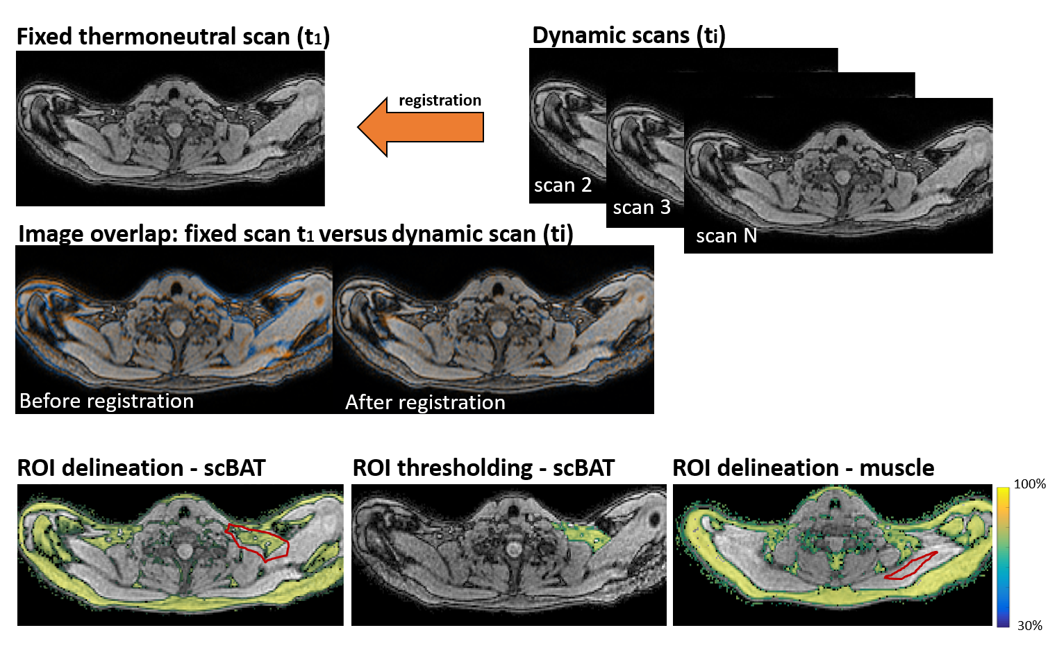
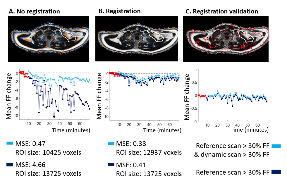
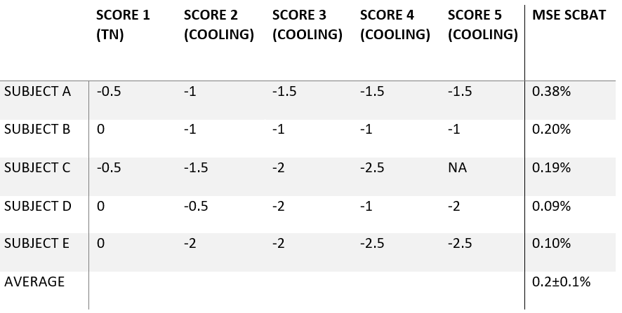
Five healthy volunteers (26.2±1.3 years; BMI 22.8±1.4 kg/m2) underwent a standardized cooling procedure for BAT activation using a water-circulating blanket. After 20 minutes at thermoneutrality (27℃), the temperature was set to 18℃ for one hour. Subjects reported thermal perceptions at thermoneutrality and every 15 minutes after cooling using a thermal perception scale ranging from cold(-3) to hot(3)11. Images were acquired at 3T using a 16-channel anterior and 12-channel posterior array and a 16-channel head-and-neck coil. Scans were acquired during the last 10 minutes at thermoneutrality and for 60 minutes during cooling using a 3D gradient-echo 12-point Dixon sequence: TR=12 ms, TE1=1.12 ms, ΔTE=0.87 ms, FA=3°, 2 mm isotropic resolution, FOV of 400×200×134 mm3 (n=2) or 400x×229×134 mm3 (n=3) with a breath-hold duration of 12 s (n=2) or 15 s (n=3), mDIXON Quant and scanner reconstructions. The acquisition time per scan was 59 s (n=2) or 1.03 m (n=3). For analysis, we co-registered first-echo magnitude images of each dynamic to the first thermoneutral scan (reference scan) using Elastix12 (Fig. 1). FF of the scBAT depot was obtained by coarsely delineating the depot on the reference scan, and non-fatty voxels below 30% FF were excluded in the reference scan and in every registered dynamic (Fig. 1). To validate the registration, we registered the reference scan of one subject to each of its dynamics, and applied the inverse transform to deform the registered reference scan back to its original coordinates with the goal of analyzing the voxel-wise FF overlap. The efficacy of registration for this subject was assessed by comparing FF dynamics with and without co-registration, and by comparing the use of FF thresholds only on the reference scan and with thresholds on all the dynamics. ROIs manually drawn in the trapezius muscle were used as a reference. Voxel-wise FF differences were calculated between the reference scan and each dynamic i: ΔFFi (x,y,z)= FFi (x,y,z) - FFTN1 (x,y,z) and averaged over both ROIs. To determine the variability, we fitted a linear line through all scBAT FF dynamics and computed the mean squared error (MSE) of residuals.Results
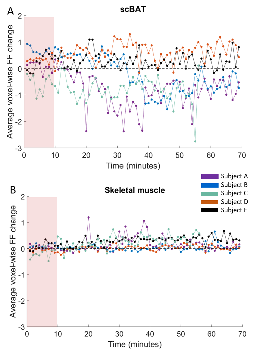
All subjects were able to adhere to the protocol. The validation and efficacy of co-registration showed a large variability in FF dynamics when applying a FF threshold on the reference scan only in non-registered data (MSE=4.67; ROI size = 13725; Fig. 2A), which was improved after thresholding voxels below 30% FF in the dynamics as well (MSE=0.47; ROI size = 10425). In registered data, this difference between single FF thresholding (MSE=0.41; ROI size=13725) and double FF thresholding (MSE=0.38; ROI size=12937) was negligible. The validation experiment showed that co-registration resulted in average voxel-wise FF changes of less than 0.5% (Fig 2C). Reported thermal scores are provided in Table 1. Voxel-wise FF differences are shown for scBAT and skeletal muscle in Figures 3A,B. We found a scBAT FF decrease in subjects A and B, whereas no changes were observed in the muscle. Subject C showed scBAT FF reductions at all time points and the last two subjects did not show any response. We found an overall MSE of 0.2±0.1% along scBAT FF dynamics (Table 1), indicating low variability.Discussion
We found that using a single FF threshold on the reference scan heavily increased the variability in scBAT FF dynamics when using non-registered data. This effect was much less apparent when using FF thresholds on all dynamics, however this restricted the ROI size due to non-overlapping FF voxels below 30% in the dynamics. Co-registration further reduced the variability and maintained a larger amount of co-located voxels. We determined an overall variability of ~0.5%, which over 60 dynamics should enable the detection of decreasing trends in cold-induced BAT FF dynamics. Cold-induced scBAT responses differed between subjects, which may be due to sub-optimal thermoneutral conditions, as not all baseline thermal scores were nonnegative.Conclusion
We showed the feasibility of obtaining 1-minute-resolution data of scBAT using breath-holds during cooling in healthy subjects. Co-registration and FF thresholding reduced motion-induced variation. Cold-induced scBAT responses varied between subjects, the reason of which is currently under investigation.Acknowledgements
References
1. Berbeé JFP, Boon MR, Khedoe PPSJ, et al. Brown fat activation reduces hypercholesterolaemia and protects from atherosclerosis development. Nat Commun. 2015;6.
2. Reber J, Willershäuser M, Karlas A, et al. Non-invasive Measurement of Brown Fat Metabolism Based on Optoacoustic Imaging of Hemoglobin Gradients. Cell Metab. 2018;27(3):689-701.e4.
3. Cypess AM, Kahn CR. Brown fat as a therapy for obesity and diabetes. Curr Opin Endocrinol Diabetes Obes. 2010;17(2):143-149.
4. Ouellet V, Labbé SM, Blondin DP, et al. Brown adipose tissue oxidative metabolism contributes to energy expenditure during acute cold exposure in humans. J Clin Invest. 2012;122(2):545-552.
5. Karampinos DC, Weidlich D, Wu M, Hu HH, Franz D. Techniques and Applications of Magnetic Resonance Imaging for Studying Brown Adipose Tissue Morphometry and Function. Handb Exp Pharmacol. 2019;251:299-324.
6. Oreskovich S, Ong F, Ahmed B, et al. Magnetic resonance imaging reveals human brown adipose tissue is rapidly activated in response to cold. J Endocr Soc. October 2019.
7. Gashi G, Madoerin P, Maushart CI, et al. MRI characteristics of supraclavicular brown adipose tissue in relation to cold-induced thermogenesis in healthy human adults. J Magn Reson Imaging. 2019;50(4):1160-1168.
8. Stahl V, Maier F, Freitag MT, et al. In vivo assessment of cold stimulation effects on the fat fraction of brown adipose tissue using DIXON MRI. J Magn Reson Imaging. 2017;45(2):369-380.
9. Lundström E, Strand R, Johansson L, Bergsten P, Ahlström H, Kullberg J. Magnetic resonance imaging cooling-reheating protocol indicates decreased fat fraction via lipid consumption in suspected brown adipose tissue. PLoS One. 2015;10(4).
10. Coolbaugh CL, Damon BM, Bush EC, Welch EB, Towse TF. Cold exposure induces dynamic, heterogeneous alterations in human brown adipose tissue lipid content. Sci Rep. 2019;9(1):13600.
11. Ashraf M. ASHRAE Handbook: Fundamentals. (2005) Inch-pound Edition ASHRAE
12. Klein S, Staring M, Murphy K, Viergever MA, Pluim JPW, elastix: a toolbox for intensity based medical image registration. IEEE Trans Med Imag. 2010; 29(1):196–205
Figures