2779
Improved image quality of liver diffusion-weighted imaging by combining compressed sensing and sensitivity encoding1Kanazawa University, Kanazawa, Japan, 2Philips Japan, Tokyo, Japan
Synopsis
Although sensitivity encoding (SENSE) technique is commonly used to reduce distortion in diffusion-weighted imaging (DWI), highly accelerated SENSE results in significantly increased noise, leading to systematic errors to the quantification of apparent diffusion coefficient. In this study, we proposed a novel method using compressed sensing combined with a highly accelerated sensitivity encoding (CS-SENSE) to reduce both the distortion and noise in liver DWI. The CS-SENSE demonstrated the higher SNR and reduced geometric distortion compared with conventional SENSE reconstruction. Liver DWI with the proposed method can improve the image quality with better lesion conspicuity.
INTRODUCTION
Diffusion-weighted imaging (DWI) of the liver with single-shot echo-planar imaging (SS-EPI) has been widely used for the detection and diagnosis of hepatic lesions.1 However, SS-EPI is highly sensitive to magnetic field inhomogeneities, causing severe geometric distortion.2 To reduce this distortion, sensitivity encoding (SENSE) technique is commonly used.3,4 Although higher SENSE acceleration factor is preferable to further improve the distortion, it results in significantly increased noise at the central parts of the image, that is, areas with a high geometry factor.5 Moreover, the presence of noise introduces systematic errors to the quantification of apparent diffusion coefficient (ADC). Therefore, to reduce both the distortion and noise in liver DWI, we proposed a novel method using compressed sensing combined with a highly accelerated SENSE (CS-SENSE).MATERIALS AND METHODS
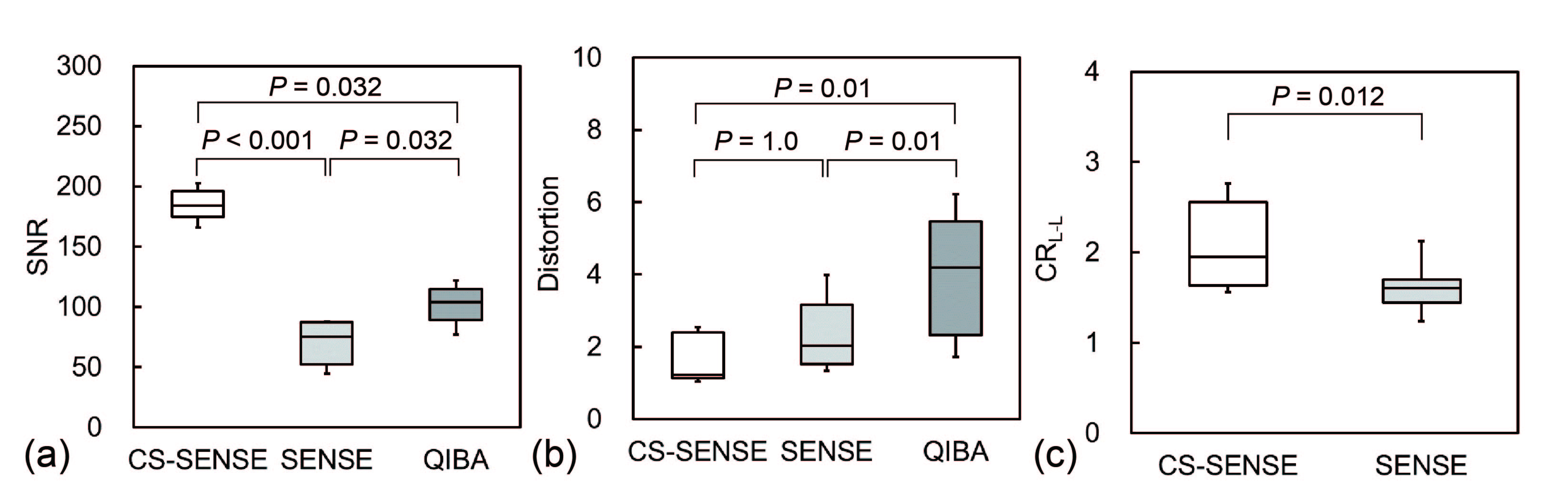
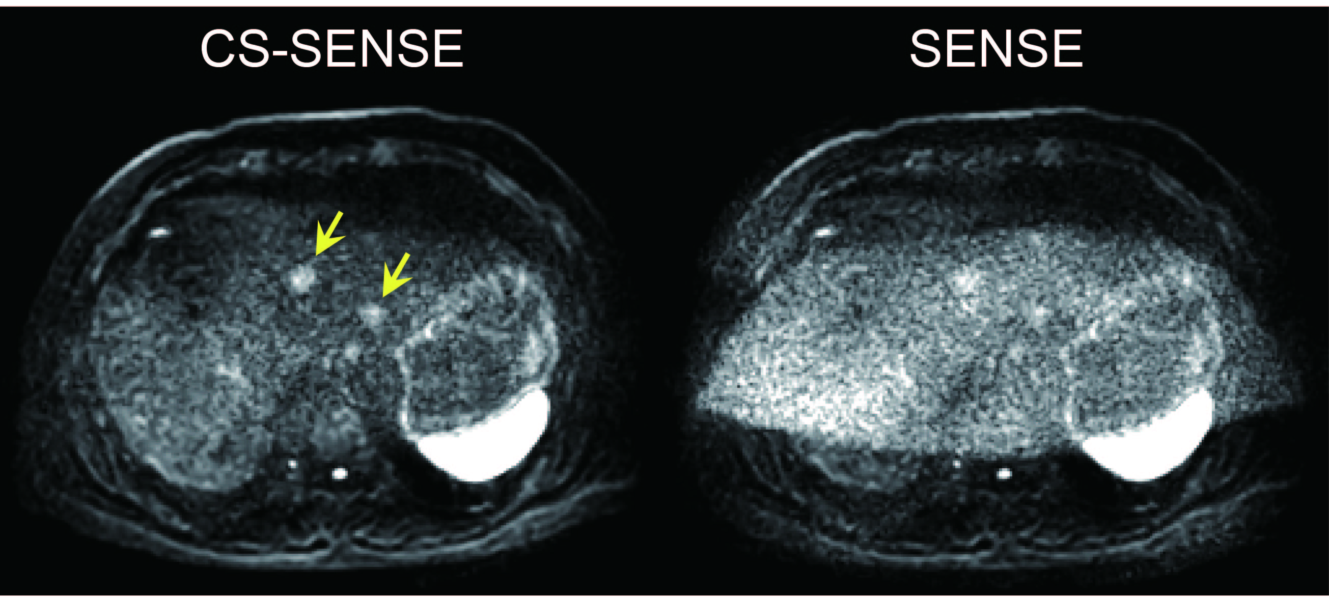
On a 3.0-T MRI, a Quantitative Imaging Biomarker Alliance (QIBA) diffusion phantom consisting of 13 vials with various concentrations of polymer polyvinylpyrrolidone in aqueous solution was scanned using diffusion-weighted (DW) SS-EPI with SENSE factor of 4 and b-values of 0, 500, and 900 s/mm2. Moreover, the scan with a QIBA standardized protocol was performed as a reference standard. To minimize the temperature dependence on ADC quantification, the phantom was filled with ice water at 0℃. Transverse DW images of the phantom were obtained with conventional SENSE and CS-SENSE reconstruction, and then the ADC maps were created. Next, we evaluated the signal-to-noise ratio (SNR) and geometric distortion of each vial in the phantom according to the National Electrical Manufacturers Association standards.6 The SNR, distortion, and ADC were compared among CS-SENSE, conventional SENSE, and QIBA using the Friedman test. Moreover, eight patients with hepatic lesions were scanned to assess the clinical utility of the proposed method. Lesion-to-liver contrast ratios (CRL-L) were measured in DW images obtained with CS-SENSE and conventional SENSE reconstruction and statistically compared using the Wilcoxon singed-rank test. A P value of <0.05 was considered statistically significant.RESULTS
In the phantom study, CS-SENSE exhibited a significantly higher SNR than QIBA protocol and conventional SENSE, and a lower distortion than QIBA (P < 0.05 for all). Moreover, the difference in ADC between CS-SENSE and QIBA was not significant. In this clinical study, CRL-L in CS-SENSE was significantly improved compared with conventional SENSE because of the substantial noise reduction (P < 0.05).CONCLUSION
A highly accelerated CS-SENSE significantly increased the SNR and reduced geometric distortion in DW SS-EPI. Liver DWI with the proposed method can improve the image quality with better lesion conspicuity.Acknowledgements
No acknowledgement found.References
1. Ni P, Lin Y, Zhong Q, et al. Technical advancements and protocol optimization of diffusion-weighted imaging (DWI) in liver. Abdom Radiol (NY) 2016;41:189-202.
2. Le Bihan D, Poupon C, Amadon A, et al. Artifacts and pitfalls in diffusion MRI. J Magn Reson Imaging 2006;24:478-488.
3. Bammer R, Keeling SL, Augustin M, et al. Improved diffusion-weighted single-shot echo-planar imaging (EPI) in stroke using sensitivity encoding (SENSE). Magn Reson Med 2001;46:548-554.
4. Pruessmann KP, Weiger M, Scheidegger MB, et al. SENSE: sensitivity encoding for fast MRI. Magn Reson Med 1999;42:952-962.
5. Dietrich O, Heiland S, Sartor K. Noise correction for the exact determination of apparent diffusion coefficients at low SNR. Magn Reson Med 2001;45:448-453.
6. National Electrical Manufacturers Association (NEMA). NEMA Standards Publication MS 1-2008; 2008.
Figures