2773
The application value of CAIPIRINHA-VIBE sequence with non-rigid 3D-registration motion correction in liver1Department of Radiology, the Second Xiangya Hospital of Central South University, Changsha, China, 2MR Scientific Marketing, Siemens Healthcare Ltd., Wuhan, China
Synopsis
This prospective study aimed to compare the image quality of T1w-VIBE sequence using generalized autocalibrating partially parallel acquisitions (GRPPA-VIBE) and controlled aliasing in parallel imaging results in higher acceleration (CAIPIRINHA-VIBE) and the spatial position consistency of CAIPIRINHA with/without non-rigid 3D-registration motion correction (MOCO) in liver MRI. The results showed that the CAIPIRINHA had better image quality than the GRPPA with respect to respiratory motion artifact suppression, liver edge sharpness and intrahepatic vascular sharpness, and the spatial position consistency of the liver using CAIPIRINHA with MOCO was significantly better than that of images without MOCO.
Introduction
Three-dimensional T1-weighted gradient recall echo volumetric interpolated breath-hold examination (VIBE) sequence is one of the most used sequences in liver magnetic resonance imaging (MRI) [1,2]. However, the conventional acceleration method, such as generalized autocalibrating partially parallel acquisitions (GRAPPA) technique, still has long scanning time about 20s and cannot meet the requirements of patients who have the problem of holding their breath[3]. The purpose of this study is to compare the image quality of VIBE sequence using a new controlled aliasing in parallel imaging results in higher acceleration (CAIPIRINHA) method and VIBE sequence using GRAPPA in liver, and to evaluate the effect of non-rigid 3D-registration motion correction (MOCO) combining with CAIPIRINHA on liver spatial location registration.Methods and Materials
The study was approved by the Ethics Committee of our hospital. A total number of 85 patients with liver MRI enhancement examination in our hospital from March 2020 to August 2020 were recruited. All the patients signed the informed consent. Liver MRI examinations of all patients were performed on a 3T MRI scanners (MAGNETOM Skyra, Siemens Healthcare, Erlangen, Germany) using 18-channel abdominal phase array coil and 32-channel Tim spinal array coil. All patients underwent pre-contrast GRAPPA-VIBE and CAIPIRINHA-VIBE breath-hold scan in the mask phase, and then underwent CAIPIRINHA-VIBE breath-hold scan in arterial phase, portal phase and delayed phase after administration. The parameters are listed in Table 1. After the scanning of four phases of CAIPIRINHA-VIBE sequence completed, 3D images without and with MOCO of each phase were automatically generated. The images quality of GRAPPA-VIBE and CAIPIRINHA-VIBE in the mask phase were scored subjectively by two physicians. The number of slices at the top of the diaphragm in the arterial phase was taken as the base slice, and the number of slices at corresponding position in the other stages subtracted with the base slice. The range of diaphragm movement in each phase was counted by + N/- N statistics.Results
The image quality and the scores of CAIPIRINHA-VIBE were significantly higher than those of GRAPPA-VIBE regarding respiratory motion artifact suppression, liver edge sharpness and intrahepatic vascular sharpness (p < 0.05), as shown in Table 2 and Figure 1. The spatial position consistency of the liver images using CAIPIRINHA-VIBE with MOCO was significantly better than those without MOCO. In the mask phase, the number of cases moving range of 0-2, 3-5 and more than 5 slices without and with MOCO changed from 65 to 83, 15 to 2 and 5 to 0, respectively. All results are summarized in Table 3, and representative images are showed in Figure 2.Discussion
The results showed that the image quality of CAIPIRINHA-VIBE sequence was superior to conventional GRAPPA-VIBE in respiratory movement artifacts, hepatic edge sharpness and intrahepatic vascular sharpness according to the image quality and the subjective scores of two radiologists. One reason maybe that CAIPIRINHA has shorter scan time and the patient can better cooperate with the scanning in a short breath-hold. The other reason maybe that CAIPIRINHA undersamples MRI data in the direction of both phase encoding and frequency encoding, and modifies aliasing artifacts during volume scanning through changing the sampling mode of phase encoding. The results were consistence with a previous study [4]. The CAIPIRINHA-VIBE with MOCO had a better position consistency of liver images than that without MOCO. This is because the liver moves in different breath-holds and the liver morphology changes during dynamic contrast-enhanced scan. The MOCO method can correct these differences. After correcting the imaging in different phases, the consistence and quantitative parameters would be improved.Conclusions
CAIPIRINHA-VIBE with MOCO can significantly improve image quality by reducing motion artifacts, so that to ensure the high consistency of spatial position of liver in different stages. In upper abdominal MRI enhancement examination, it can be used instead of conventional GRAPPA-VIBE sequence, especially in patients with poor breath-hold ability.Acknowledgements
No acknowledgement found.References
[1]Ba-Ssalamah A., Baroud S. & Bastati N. et al., "MR Imaging of Benign Focal Liver Lesions," Magnetic Resonance Imaging Clinics of North America, Vol.18, No.3(2010).
[2]Yu M. H., Lee J. M. & Yoon J. H. et al., "Clinical application of controlled aliasing in parallel imaging results in a higher acceleration (CAIPIRINHA)-volumetric interpolated breathhold (VIBE) sequence for gadoxetic acid-enhanced liver MR imaging," J Magn Reson Imaging, Vol.38, No.5(2013), pp.1020-1026.
[3]Shin P. Y., Hee L. C. & Seong K. I. et al., "Usefulness of controlled aliasing in parallel imaging results in higher acceleration in gadoxetic acid-enhanced liver magnetic resonance imaging to clarify the hepatic arterial phase.," Investigative radiology, Vol.49, No.3(2014).
[4]Riffel P., Attenberger U. I. & Kannengiesser S. et al., "Highly accelerated T1-weighted abdominal imaging using 2-dimensional controlled aliasing in parallel imaging results in higher acceleration: a comparison with generalized autocalibrating partially parallel acquisitions parallel imaging.," Investigative radiology, Vol.48, No.7(2013).
Figures

Table 1. Scanning parameters of GRAPPA-VIBE and CAIPIRINHA-VIBE sequences.
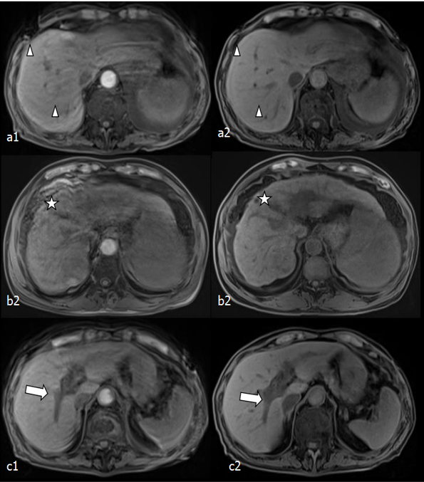
Figure 1. a1-c1 and a2-c2 are the images using the GRAPPA-VIBE and the MOCO-CAIPI-VIBE sequences, respectively. a2-c2 are better in respiratory movement artifacts (triangles), definition (star), and the display of intrahepatic vessels (arrows).

Table 2. Qualitative Analysis of GRAPPA-VIBE and CAIPIRINHA-VIBE sequences. All values are mean ±standard deviation of the scores.

Table 3. The number of moving slices in different phase.
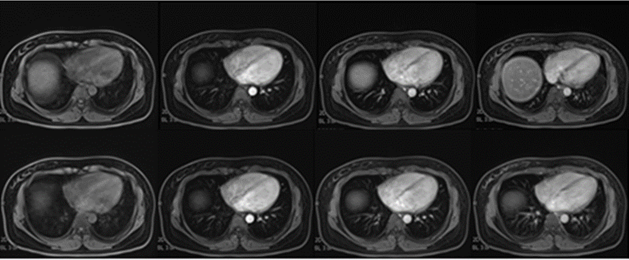
Figure 2. The images using CAIPIRINHA-VIBE without MOCO (the upper row) and with MOCO (the lower row) in four phases, including mask, arterial, portal, delayed phases (from left to right).